. 2016 Feb 12:7:10731.
doi: 10.1038/ncomms10731.

Tripartite assembly of RND multidrug efflux pumps

Affiliations

Tripartite assembly of RND multidrug efflux pumps

Laetitia Daury et al. Nat Commun. .

Abstract

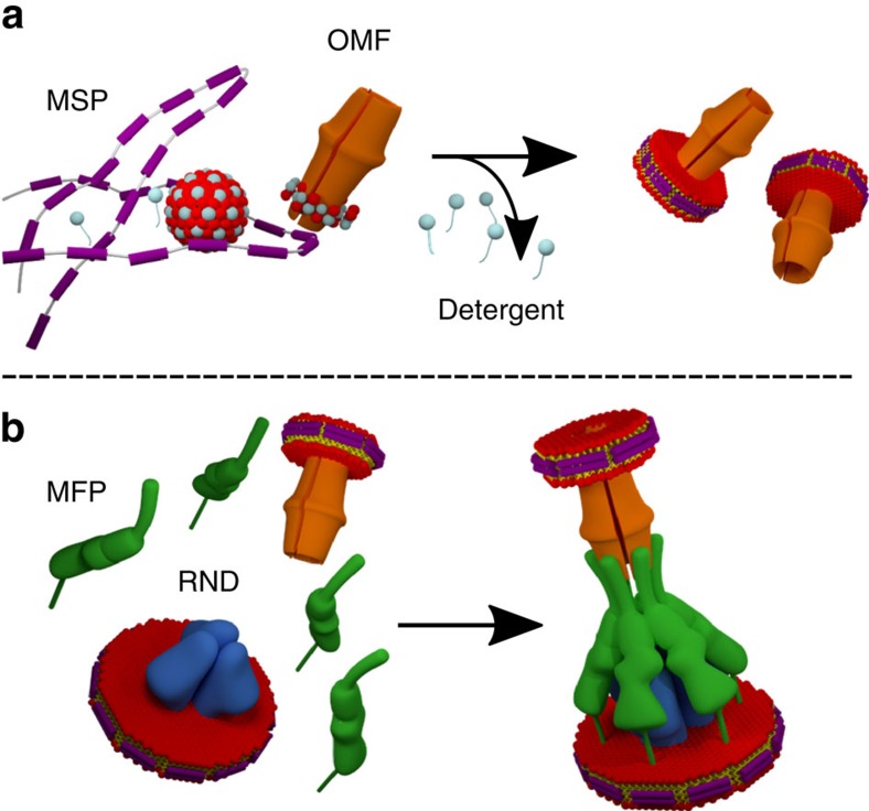
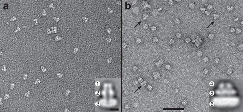
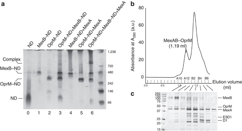
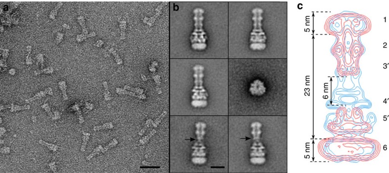
Tripartite multidrug efflux systems of Gram-negative bacteria are composed of an inner membrane transporter, an outer membrane channel and a periplasmic adaptor protein. They are assumed to form ducts inside the periplasm facilitating drug exit across the outer membrane. Here we present the reconstitution of native Pseudomonas aeruginosa MexAB-OprM and Escherichia coli AcrAB-TolC tripartite Resistance Nodulation and cell Division (RND) efflux systems in a lipid nanodisc system. Single-particle analysis by electron microscopy reveals the inner and outer membrane protein components linked together via the periplasmic adaptor protein. This intrinsic ability of the native components to self-assemble also leads to the formation of a stable interspecies AcrA-MexB-TolC complex suggesting a common mechanism of tripartite assembly. Projection structures of all three complexes emphasize the role of the periplasmic adaptor protein as part of the exit duct with no physical interaction between the inner and outer membrane components.


Figures

Figure 1
Figure 1. Tripartite assembly based on RND and OMF inserted into nanodiscs.
(a) After detergent removal, the integral membrane proteins are reconstituted into a small lipid bilayer wrapped by two MSPs (purple) forming the nanodisc. Lipids are red/yellow and detergent is grey. (b) Self-assembly of RND (blue) and OMF (orange) in nanodiscs in the presence of native lipid-modified MFP (green) leading to the tripartite complex in lipid membrane.
Figure 2
Figure 2. TEM observations of OprM and MexB reconstituted into nanodiscs.
(a) Field of view of OprM–ND showing side views of isolated molecules. The average image (inset) reveals characteristic features: The OprM β-barrel in the ND (1) and the OprM periplasmic domain composed of the equatorial domain (2) and the tip of the α-barrel (3) protruding from the ND. (b) Field of view of MexB-ND showing isolated molecules. Black arrows indicate side views. On the average image (inset), a side view of MexB exhibits the periplasmic part organized in two layers (funnel (4) and porter (5) domains) protruding from the ND (6). Scale bars, 50 nm and 5 nm for the inset. a.u., arbitrary unit.
Figure 3
Figure 3. Native PAGE analysis and purification of the tripartite MexAB–OprM assembly.
(a) Electrophoretic mobility shift assay of individual and mixed components. OprM–ND and MexB–ND were mixed in the presence and in the absence of MexA. An extra band was observed when the three components were present in the sample. Lane 0, ND; lane 1, MexB–ND; lane 2, OprM–ND; lane 3, MexB–ND and OprM–ND; lane 4, MexB–ND and MexA; lane 5, OprM–ND and MexA; lane 6, MexB–ND, OprM–ND, and MexA. Proteins were separated by native PAGE and stained with PlusOne Silver Staining Kit. (b) Analytical size-exclusion chromatography (SEC) analysis of the mixed components. (c) SDS–PAGE analysis of the indicated SEC peak fractions. The molecular mass of each marker protein (in kilodalton) is indicated on the right (a) and on the left (b).
Figure 4
Figure 4. TEM analysis of tripartite MexAB–OprM assembly.
(a) Field of view revealing elongated complexes when OprM-ND and MexB-ND were mixed in the presence of MexA. Scale bar, 30 nm. (b) Gallery of five class average side views of a 33-nm-long tripartite MexAB–OprM complex delineated by two nanodiscs (157, 192, 185, 99, 151 images, respectively) and one top view class average (56 images). Lower row, two average classes of atypical complexes showing faint contacts between OprM and MexAB (black arrows). Scale bar, 10 nm. (c) Isocontours of MexB–ND and OprM–ND (red) overlaid on isocontours of tripartite complex (blue). Characteristic features are displayed: OprM β-barrel and ND (1); equatorial domain (2), tip of α barrel (3′), MexB funnel (4′) and porter (5′) domains anchored to ND (6). The remaining blue densities correspond to MexA that linked OprM to MexB and interacts with the domains marked with (′). The tripartite assembly ND (6) has a smaller size compared with ND of MexB probably because of the detergent carried with MexA that may extract some lipids.
Figure 5
Figure 5. TEM analysis of tripartite AcrAB–TolC and AcrA–MexB–TolC assemblies.
(a) Field of view revealing purified AcrAB–TolC assemblies. Scale bar, 30 nm. (b) Gallery of one top view class average (137 images) and four class average side views of a 33-nm-long tripartite AcrAB–TolC complex (223, 119, 196, 178 images, respectively) and. (c) Enlarged average image of tripartite AcrAB–TolC assembly. (d) Isocontours of tripartite AcrAB–TolC assembly (red) overlaid on isocontours of tripartite MexAB–OprM assembly (blue). (e) An average of tripartite AcrA–MexB–TolC assembly. Scale bar, 10 nm for c,d,e.

Similar articles

Cited by

References

    1. May M. Drug development: time for teamwork. Nature 509, S4–S5 (2014). - PubMed
    1. Li X.-Z., Plésiat P. & Nikaido H. The challenge of efflux-mediated antibiotic resistance in Gram-negative bacteria. Clin. Microbiol. Rev. 28, 337–418 (2015). - PMC - PubMed
    1. Nikaido H. Multidrug resistance in bacteria. Annu. Rev. Biochem. 78, 119–146 (2009). - PMC - PubMed
    1. Koronakis V., Sharff A., Koronakis E., Luisi B. & Hughes C. Crystal structure of the bacterial membrane protein TolC central to multidrug efflux and protein export. Nature 405, 914–919 (2000). - PubMed
    1. Mikolosko J., Bobyk K., Zgurskaya H. I. & Ghosh P. Conformational flexibility in the multidrug efflux system protein AcrA. Structure 14, 577–587 (2006). - PMC - PubMed

Publication types

MeSH terms

Substances