. 2016 Apr 1;291(14):7727-41.
doi: 10.1074/jbc.M115.701540. Epub 2016 Feb 11.

Structural Studies of Lipopolysaccharide-defective Mutants from Brucella melitensis Identify a Core Oligosaccharide Critical in Virulence

Affiliations

Structural Studies of Lipopolysaccharide-defective Mutants from Brucella melitensis Identify a Core Oligosaccharide Critical in Virulence

Carolina Fontana et al. J Biol Chem. .

Abstract

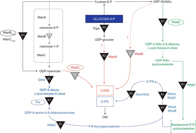
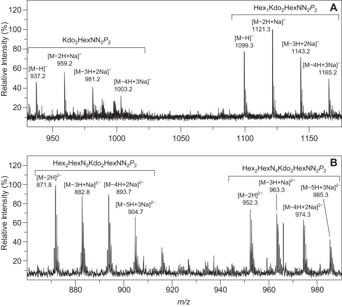
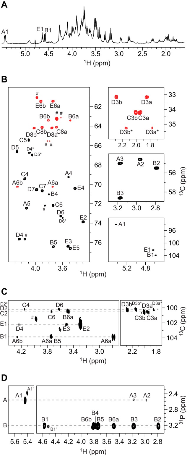
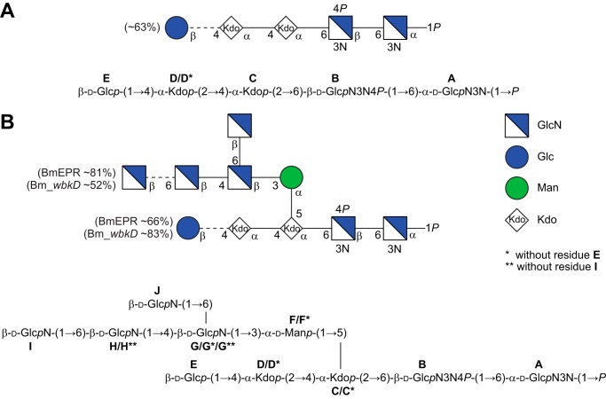
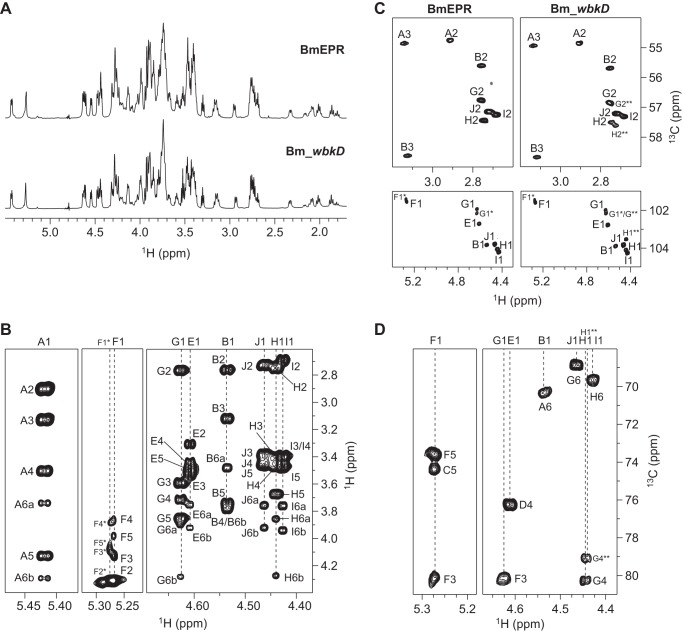
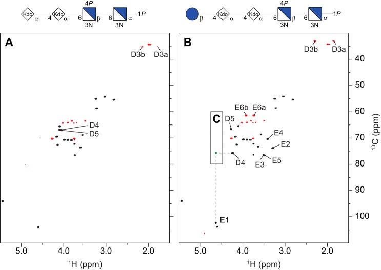
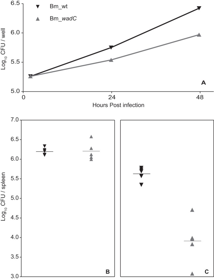
The structures of the lipooligosaccharides fromBrucella melitensismutants affected in the WbkD and ManBcoreproteins have been fully characterized using NMR spectroscopy. The results revealed that disruption ofwbkDgives rise to a rough lipopolysaccharide (R-LPS) with a complete core structure (β-d-Glcp-(1→4)-α-Kdop-(2→4)[β-d-GlcpN-(1→6)-β-d-GlcpN-(1→4)[β-d-GlcpN-(1→6)]-β-d-GlcpN-(1→3)-α-d-Manp-(1→5)]-α-Kdop-(2→6)-β-d-GlcpN3N4P-(1→6)-α-d-GlcpN3N1P), in addition to components lacking one of the terminal β-d-GlcpN and/or the β-d-Glcpresidues (48 and 17%, respectively). These structures were identical to those of the R-LPS fromB. melitensisEP, a strain simultaneously expressing both smooth and R-LPS, also studied herein. In contrast, disruption ofmanBcoregives rise to a deep-rough pentasaccharide core (β-d-Glcp-(1→4)-α-Kdop-(2→4)-α-Kdop-(2→6)-β-d-GlcpN3N4P-(1→6)-α-d-GlcpN3N1P) as the major component (63%), as well as a minor tetrasaccharide component lacking the terminal β-d-Glcpresidue (37%). These results are in agreement with the predicted functions of the WbkD (glycosyltransferase involved in the biosynthesis of the O-antigen) and ManBcoreproteins (phosphomannomutase involved in the biosynthesis of a mannosyl precursor needed for the biosynthesis of the core and O-antigen). We also report that deletion ofB. melitensis wadCremoves the core oligosaccharide branch not linked to the O-antigen causing an increase in overall negative charge of the remaining LPS inner section. This is in agreement with the mannosyltransferase role predicted for WadC and the lack of GlcpN residues in the defective core oligosaccharide. Despite carrying the O-antigen essential inB. melitensisvirulence, the core deficiency in thewadCmutant structure resulted in a more efficient detection by innate immunity and attenuation, proving the role of the β-d-GlcpN-(1→6)-β-d-GlcpN-(1→4)[β-d-GlcpN-(1→6)]-β-d-GlcpN-(1→3)-α-d-Manp-(1→5) structure in virulence.

Keywords: Brucella melitensis; Gram-negative bacteria; WadC; glycosyltransferase; lipopolysaccharide (LPS); mutant; nuclear magnetic resonance (NMR).


Figures

FIGURE 1.
FIGURE 1.
Proposed pathways involved in the biosynthesis of the S-LPS of B. melitensis. Overview of the pathways involved in the biosynthesis of the S-LPS of B. melitensis (adapted from Ref. 10). The steps leading to N-formylperosamine synthesis and to its polymerization are indicated in blue, and those leading to bactoprenol priming for N-formylperosamine polymerization are depicted in green. Subsequently, the O-PS is translocated to the periplasm by the Wzm/Wzt ABC transporter (also in blue) and ligated to the core oligosaccharide that results from the pathways marked in red. The LPS phenotypes obtained by disrupting the different steps (indicated by black triangles) are annotated (R1, R2, or R3). A white-filled triangle (GDP-mannose pathway) indicates a mutation that does not generate an R phenotype, and gray triangles marked with S (middle) indicate a mutation that, while blocking the synthesis of a core lateral branch, does not prevent O-PS linkage to the core. Enzymes disrupted in this study (WadC, Per, WbkD, and ManBcore) are indicated with an ellipse.
FIGURE 2.
FIGURE 2.
Mass spectra of deacylated LOS. Selected regions of the high resolution mass spectra of the deacylated core oligosaccharides of Bm_manBcore (A) and Bm_wbkD (B) recorded in negative ion mode. The clusters of pseudo-molecular ions originating from the two major components of each sample are annotated (see Table 2). Note that the peaks observed in A and B correspond to singly and doubly negatively charged ion species, respectively.
FIGURE 3.
FIGURE 3.
NMR spectra of the deacylated LOS from Bm_manBcore. A, selected region of the diffusion-filtered 1H NMR spectrum. B, selected regions of the multiplicity-edited 1H,13C HSQC spectrum showing the anomeric region (right bottom), the region for the nitrogen-bearing carbons (right middle), the region for the 3-deoxy-groups of the Kdo residues (right top), and the region for the ring atoms and those from hydroxymethyl groups (left) in which the cross-peaks from the latter appear in red. C, selected region of the 1H,13C HMBC spectrum showing intra- and inter-residue correlations from anomeric carbons. D, selected region of the 1H,31P-hetero TOCSY spectrum (τm = 92 ms) showing correlations from the phosphate groups in residues A and B. Signals from impurities of lower molecular mass than the LOS oligosaccharides of B. melitensis are indicated by the hash symbol.
FIGURE 4.
FIGURE 4.
Structures of the deacylated LOS from Bm_manBcore, Bm_wbkD, and BmEPR. A, structure of the penta- and tetra-saccharides in CFG format (top) and standard nomenclature (bottom) obtained by deacylation of the LOS from Bm_manBcore. B, structure of the deca-, nona-, and octasaccharides in CFG format (top) and standard nomenclature (bottom) obtained by deacylation of the LOS from BmEPR and Bm_wbkD. The ratio of the different oligosaccharides present in the samples was estimated by integration of selected cross-peaks in the multiplicity-edited 1H,13C HSQC spectrum; the D3b and D3b* resonances were used to estimate the ratio of oligosaccharides with and without residue E, whereas the G4 and G4** resonances were used to estimate the ratio of oligosaccharides with and without residue I.
FIGURE 5.
FIGURE 5.
NMR spectra of the deacylated LOS from Bm_wbkD and BmEPR. A, comparison of the diffusion-filtered 1H NMR spectra of the deacylated LOS from BmEPR and Bm_wbkD (top and bottom, respectively). B, selected regions of the 1H,1H TOCSY (τm = 100 ms) of deacylated LOS from BmEPR showing correlations from anomeric protons. C, comparison of selected regions of the multiplicity-edited 1H,13C HSQC spectrum of the deacylated LOS from BmEPR and Bm_wbkD (left and right, respectively), showing part of the anomeric region (bottom) and the region for the nitrogen-bearing carbons (top). D, selected regions of the 1H,13C HMBC spectrum of the deacylated LOS from Bm_wbkD showing intra- and inter-residue correlations from anomeric protons.
FIGURE 6.
FIGURE 6.
NMR spectra of the deacylated LOS from Bm_wadC_per. Comparison of the multiplicity-edited 1H,13C HSQC spectra (black/red) of the tetrasaccharide (A) and pentasaccharide (B) (OS1 and OS2, respectively) isolated from Bm_wadC_per; the cross-peaks with significant chemical shift differences between the two oligosaccharides are annotated. The structures (in CFG format) of the respective oligosaccharides are shown on the top of each spectra. C, overlay of a selected region of the 1H,13C HMBC spectrum of the OS2 showing an interglycosidic heteronuclear long range correlation (green) between residues D and E.
FIGURE 7.
FIGURE 7.
B. melitensis WadC mutants carry a core defect that does not affect the linkage to the O-PS. SDS-PAGE and Western blot analysis with anti-O-chain monoclonal antibody Cby-33H8 of LPS SDS-proteinase K extracts from Bm_wt (B. melitensis 16M wild type) and Bm_wadC (B. melitensis 16MΔwadC mutant).
FIGURE 8.
FIGURE 8.
B. melitensis core lateral branch shields bacterial surface negatively charged groups (inner core and lipid A). The figure presents ζ potential measurements of Bm_per (no LPS O-PS and complete core oligosaccharide), Bm_wadC_per (no LPS O-PS and defective core oligosaccharide) in comparison with B. melitensis 16M (wild type strain; Bm_wt). Each bar represents the means ± S.E. of 10 measurements of one representative experiment.
FIGURE 9.
FIGURE 9.
B. melitensis mutants defective in the core lateral branch are attenuated in dendritic cells and in mice. Bone marrow-derived dendritic cells were infected with B. melitensis 16M (wild type strain; Bm_wt) or B. melitensis 16MΔwadC (Bm_wadC), and CFU were measured at the indicated times (each point represents the means ± S.E. of triplicate wells of a representative experiment) (A). Groups of five mice were infected with 5 × 10⁴ CFU of Bm_wt (▾) or Bm_wadC (▴) and CFU/spleen determined at 2 (B) and 8 (C) weeks.
FIGURE 10.
FIGURE 10.
B. melitensis wadC LPS triggers dendritic cell activation and maturation. Mouse BMDCs were stimulated for 24 h with medium or purified LPS from B. melitensis 16M (wild type strain; Bm_wt), B. melitensis 16MΔwadC (Bm_wadC), or E. coli (O55:B5), all administered at an equivalent molarity (0.25 μm). IFN-γ, TNF-α, IL-6, and IL-12p40 secretion levels in culture supernatant were determined by ELISA (A). Surface levels of MHC-II, CD86, CD80, and CD40 and were measured by flow cytometry (B). Data are presented as the means ± S.E. of at least five independent experiments.

References

    1. Dean A. S., Crump L., Greter H., Schelling E., and Zinsstag J. (2012) Global burden of human brucellosis: a systematic review of disease frequency. PLoS Negl. Trop. Dis. 6, e1865. - PMC - PubMed
    1. McDermott J., Grace D., and Zinsstag J. (2013) Economics of brucellosis impact and control in low-income countries. Rev. Sci. Tech. 32, 249–261 - PubMed
    1. Ariza J. (1999) Brucellosis: an update. The perspective from the Mediterranean basin. Rev. Med. Micriobiol. 10, 125–135
    1. Raetz C. R., and Whitfield C. (2002) Lipopolysaccharide endotoxins. Annu. Rev. Biochem. 71, 635–700 - PMC - PubMed
    1. Valvano M. A., Furlong S. E., and Patel K. B. (2011) in Bacterial Lipopolysaccharides (Knirel Y. A., and Valvano M. A., eds) pp. 275–310, Springer-Verlag, Wien

MeSH terms

LinkOut - more resources