. 2016 Apr 8;291(15):7858-67.
doi: 10.1074/jbc.M115.706861. Epub 2016 Feb 10.

The Platelet Integrin αIIbβ3 Differentially Interacts with Fibrin Versus Fibrinogen

Affiliations

The Platelet Integrin αIIbβ3 Differentially Interacts with Fibrin Versus Fibrinogen

Rustem I Litvinov et al. J Biol Chem. .

Abstract

Fibrinogen binding to the integrin αIIbβ3 mediates platelet aggregation and spreading on fibrinogen-coated surfaces. However,in vivoαIIbβ3 activation and fibrinogen conversion to fibrin occur simultaneously, although the relative contributions of fibrinogenversusfibrin to αIIbβ3-mediated platelet functions are unknown. Here, we compared the interaction of αIIbβ3 with fibrin and fibrinogen to explore their differential effects. A microscopic bead coated with fibrinogen or monomeric fibrin produced by treating the immobilized fibrinogen with thrombin was captured by a laser beam and repeatedly brought into contact with surface-attached purified αIIbβ3. When αIIbβ3-ligand complexes were detected, the rupture forces were measured and displayed as force histograms. Monomeric fibrin displayed a higher probability of interacting with αIIbβ3 and a greater binding strength. αIIbβ3-fibrin interactions were also less sensitive to inhibition by abciximab and eptifibatide. Both fibrinogen- and fibrin-αIIbβ3 interactions were partially inhibited by RGD peptides, suggesting the existence of common RGD-containing binding motifs. This assumption was supported using the fibrin variants αD97E or αD574E with mutated RGD motifs. Fibrin made from a fibrinogen γ'/γ' variant lacking the γC αIIbβ3-binding motif was more reactive with αIIbβ3 than the parent fibrinogen. These results demonstrate that fibrin is more reactive with αIIbβ3 than fibrinogen. Fibrin is also less sensitive to αIIbβ3 inhibitors, suggesting that fibrin and fibrinogen have distinct binding requirements. In particular, the maintenance of αIIbβ3 binding activity in the absence of the γC-dodecapeptide and the α-chain RGD sequences suggests that the αIIbβ3-binding sites in fibrin are not confined to its known γ-chain and RGD motifs.

Keywords: adhesion; fibrin; fibrinogen; integrin; platelet.


Figures

FIGURE 1.
FIGURE 1.
Diagram of the fibrinogen molecule with the Aα (blue), Bβ (red), and γ (green) chains indicated. Putative αIIbβ3 binding sites are denoted with the color of the corresponding fibrinogen chain. The location of the sequences Aα1–26, Bβ1–57, γ395–411, and Aα221–610 were added computationally to the fibrinogen crystal structure (Protein Data Bank code 3GHG).
FIGURE 2.
FIGURE 2.
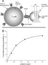
A, schematic representation of the model system used to study bimolecular interactions between αIIbβ3 and fibrinogen or fibrin. A latex bead coated with fibrinogen or fibrin is trapped by a focused laser beam and oscillated toward or away from the silica pedestal coated with αIIbβ3, touching it repeatedly. When the surface-bound αIIbβ3 and fibrinogen or fibrin monomer molecules interact, a tensile (rupture) force is generated as the bead moves to the right. B, cumulative probability of αIIbβ3-fibrinogen interactions plotted against the fibrinogen concentration in the binding solution that determines the density of fibrinogen on the surface of the coated latex bead. The data shown are representative of three experiments.
FIGURE 3.
FIGURE 3.
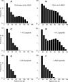
Rupture force spectra for αIIbβ3 binding to either wild-type fibrinogen or wild-type fibrin in the absence and presence of inhibitors. A, αIIbβ3 binding to immobilized wild-type fibrinogen. B, αIIbβ3 binding to immobilized wild-type fibrin monomer. Fibrin monomer was produced by treating fibrinogen-coated latex beads with thrombin. C–F, binding interactions shown in A and B repeated in the presence of 1 mm γC-dodecapeptide (H12) (C and D) or 1 mm cyclic RGD (cRGD) peptide (E and F). As described under “Experimental Procedures,” data were collected from not less than 10 pedestal bead pairs collected in three or more experiments. The histograms shown in the figure resulted from 13,580 contacts in A; 14,356 contacts in B; 6,334 contacts in C; 10,367 contacts in D; 9,564 contacts in E, and 7,867 contacts in F.
FIGURE 4.
FIGURE 4.
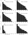
A–D, rupture force spectra for αIIbβ3 binding to recombinant fibrinogen or fibrin containing either two γ′ chains (A and B) or two γA chains (C and D). Fibrin monomers were generated as described in Fig. 3. E and F, binding interactions shown in C and D in the presence of 1 mm H12. The data were collected as described under “Experimental Procedures” and in the legend to Fig. 3. The histograms shown in the figure were generated from 11,356 contacts in A; 12,563 contacts in B; 6,334 contacts in C; 9,367 contacts in D; 6,856 contacts in E; and 9,564 contacts in F.
FIGURE 5.
FIGURE 5.
Rupture force spectra for αIIbβ3 binding to recombinant fibrinogen and fibrin in which the RGDS motif at (A)α572–575 was mutated to RGES [(A)αD574E]. A, αIIbβ3 binding to AαD574E fibrinogen. B, αIIbβ3 binding to AαD574E fibrin. C and D, αIIbβ3 binding to (A)αD574E fibrinogen and fibrin in the presence of 1 mm H12. The data were collected as described under “Experimental Procedures” and in the legend to Fig. 3. The histograms shown in the figure were generated from 10,223 contacts in A; 11,445 contacts in B; 7,357 contacts in C; and 8,289 contacts in D.
FIGURE 6.
FIGURE 6.
Rupture force spectra for αIIbβ3 binding to recombinant fibrinogen and fibrin in which the RGDF motif at (A)α95–98 was mutated to RGEF [(A)αD97E]. A, αIIbβ3 binding to AαD97E fibrinogen. B, αIIbβ3 binding to αD97E fibrin. C and D, αIIbβ3 binding to (A)αD97E fibrinogen and fibrin in the presence of 1 mm H12. The data were collected as described under “Experimental Procedures” and in the legend to Fig. 3. The histograms shown in the figure were generated from 11,654 contacts in A; 11,114 contacts in B; 6,244 contacts in C; and 7,075 contacts in D.
FIGURE 7.
FIGURE 7.
Cumulative probability of αIIbβ3-fibrinogen and αIIbβ3-fibrin binding plotted against the concentrations of abciximab (A) and eptifibatide (B) added to the reaction chamber. The plots were generated by fitting the experimental results to the exponential functions shown in the figure. The results shown are the means and standard deviations of three experiments.
FIGURE 8.
FIGURE 8.
Cartoon illustrating the potential role of fibrin in platelet aggregation. Platelet aggregation initially begins with fibrinogen (Fg) binding to αIIbβ3 but quickly evolves into αIIbβ3 interaction with monomeric, oligomeric, or polymeric fibrin (Fn) as fibrinogen is converted to fibrin by thrombin generated at or near the surface of the activated platelets.

References

    1. Bennett J. S. (2001) Platelet-fibrinogen interactions. Ann. N.Y. Acad. Sci. 936, 340–354 - PubMed
    1. Dai A., Ye F., Taylor D. W., Hu G., Ginsberg M. H., and Taylor K. A. (2015) The structure of a full-length membrane-embedded integrin bound to a physiological ligand. J. Biol. Chem. 290, 27168–27175 - PMC - PubMed
    1. Collet J. P., Montalescot G., Lesty C., and Weisel J. W. (2002) A structural and dynamic investigation of the facilitating effect of glycoprotein IIb/IIIa inhibitors in dissolving platelet-rich clots. Circ. Res. 90, 428–434 - PubMed
    1. Carr M. E., Jr. (2003) Development of platelet contractile force as a research and clinical measure of platelet function. Cell Biochem. Biophys. 38, 55–78 - PubMed
    1. Hantgan R. R., and Mousa S. A. (1998) Inhibition of platelet-mediated clot retraction by integrin antagonists. Thromb. Res. 89, 271–279 - PubMed

Publication types

MeSH terms

Associated data

LinkOut - more resources