Review
. 2016 May;34(5):394-407.
doi: 10.1016/j.tibtech.2016.01.002. Epub 2016 Feb 9.

Gelatin-Methacryloyl Hydrogels: Towards Biofabrication-Based Tissue Repair

Affiliations
Review

Gelatin-Methacryloyl Hydrogels: Towards Biofabrication-Based Tissue Repair

Barbara J Klotz et al. Trends Biotechnol. 2016 May.

Abstract

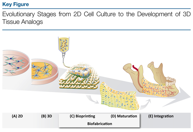
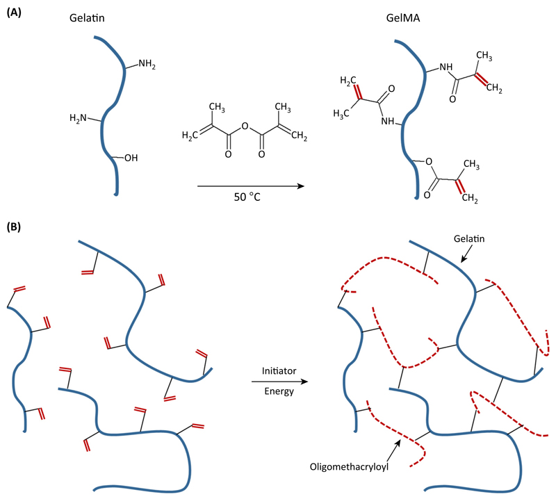
Research over the past decade on the cell-biomaterial interface has shifted to the third dimension. Besides mimicking the native extracellular environment by 3D cell culture, hydrogels offer the possibility to generate well-defined 3D biofabricated tissue analogs. In this context, gelatin-methacryloyl (gelMA) hydrogels have recently gained increased attention. This interest is sparked by the combination of the inherent bioactivity of gelatin and the physicochemical tailorability of photo-crosslinkable hydrogels. GelMA is a versatile matrix that can be used to engineer tissue analogs ranging from vasculature to cartilage and bone. Convergence of biological and biofabrication approaches is necessary to progress from merely proving cell functionality or construct shape fidelity towards regenerating tissues. GelMA has a critical pioneering role in this process and could be used to accelerate the development of clinically relevant applications.

Keywords: biofabrication; gelatin-methacryloyl; hydrogel; photo-crosslinking; regenerative medicine; tissue engineering.


Figures

Figure 1
Figure 1
(A) 2D cell culture on plastic; (B) 3D cell culture inside hydrogel constructs; (C) bioprinting of 3D constructs; (D) biological maturation of the 3D bioprinted construct forming a tissue analog; and (E) implantation and integration of the tissue analog into the defect site.
Figure 2
Figure 2. Gelatin-Methacryloyl (GelMA) Synthesis and Hydrogel Formation.
(A) Reaction of methacrylic anhydride with amine and hydroxyl groups on gelatin gives rise to gelMA macromers. (B) Upon generation of a free radical (e.g., by light exposure in the presence of a photoinitiator), the methacrylamide and methacrylate side groups on the gelMA chains polymerize via radical addition-type polymerization to yield a network of gelatin chains connected through short polymethacryloyl chains.
Figure 3
Figure 3. Categories of Gelatin-Methacryloyl (GelMA)-Based Biofabrication-Related Techniques and their Generated Constructs.
(A) Preparation of cell-laden microspheres by microfluidics (scale bar = 30 μm). (B) Stereolithographic fabrication of pyramid-shaped scaffolds by spatially controlled light-initiated crosslinking of a gelMA macromer-cell mixture in a computer-controlled platform. By using this approach, cells are encapsulated while building the construct. Encapsulated cells stained for actin expression (scale bar = 100 μm). (C) Computer-controlled robotic dispensing of cell-laden gelMA to build a 3D construct, for instance, a bioprinted analog of the distal femur from a human knee. Reproduced, with permission, from [92] (A), [42] (B), and (upper picture) [93] and (lower picture) [45] (C). Abbreviation: UV, ultraviolet.
Figure 4
Figure 4. Examples of Combining Gelatin-Methacryloyl (GelMA) with Different Materials to Obtain Tissue-Specific Functionalities.
(A) Mechanical reinforcement of hydrogels by combination with electrospun box structures that form a macroscopic network structure (scale bar = 1 mm). (B) (i) Providing gelMA with electrical conductivity by the addition of carbon nanotubes to the bulk hydrogel. (ii) The cardiomyocyte-seeded composite showed improved contraction behavior, resulting in movement of the construct of about 2.5 mm. (C) Optimizing gelMA by addition of cartilage-derived matrix particles [1.5% (w/v)] to the hydrogel (scale bar = 500 μm). Mesenchymal stromal cells (MSCs) produced pronounced cartilage-specific matrix, of GAGs and collagen type II. compared with non-laden gelMA (scale bar = 200 μm). Reproduced, with permission, from [51] (A), [55] (B, i), [58] (B, ii), and [50] (C). Abbreviation: ECM, extracellular matrix.

Similar articles

Cited by

References

    1. Pampaloni F, et al. The third dimension bridges the gap between cell culture and live tissue. Nat Rev Mol Cell Biol. 2007;8:839–845. - PubMed
    1. Ehrbar M, et al. Enzymatic formation of modular cell-instructive fibrin analogs for tissue engineering. Biomaterials. 2007;28:3856–3866. - PubMed
    1. Benton JA, et al. Photocrosslinking of gelatin macromers to synthesize porous hydrogels that promote valvular interstitial cell function. Tissue Eng Part A. 2009;15:3221–3230. - PMC - PubMed
    1. Bertassoni LE, et al. Hydrogel bioprinted microchannel networks for vascularization of tissue engineering constructs. Lab Chip. 2014;14:2202–2211. - PMC - PubMed
    1. Occhetta P, et al. VA-086 methacrylate gelatine photopolymerizable hydrogels: a parametric study for highly biocompatible 3D cell embedding. J Biomed Mater Res Part A. 2014;103:2109–2117. - PubMed

Publication types