. 2016 Oct;34(10):1716-1724.
doi: 10.1002/jor.23192. Epub 2016 Feb 26.

Nutlin-3 treatment spares cisplatin-induced inhibition of bone healing while maintaining osteosarcoma toxicity

Affiliations

Nutlin-3 treatment spares cisplatin-induced inhibition of bone healing while maintaining osteosarcoma toxicity

Kimo C Stine et al. J Orthop Res. 2016 Oct.

Abstract

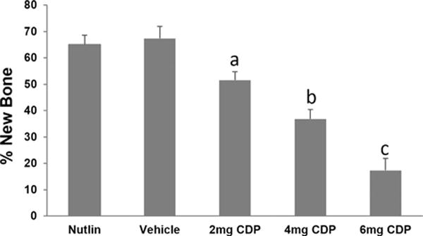
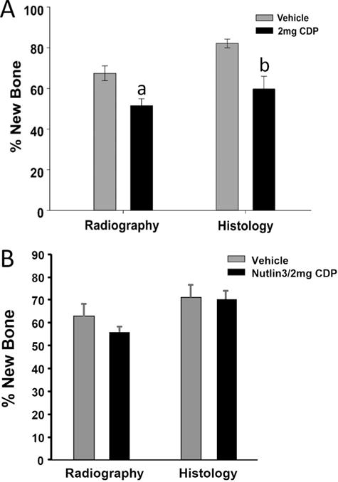
The majority of Osteosarcoma (OS) patients are treated with a combination of chemotherapy, resection, and limb salvage protocols. These protocols include distraction osteogenesis (DO), which is characterized by direct new bone formation. Cisplatin (CDP) is extensively used for OS chemotherapy and recent studies, using a mouse DO model, have demonstrated that CDP has profound negative effects on bone repair. Recent oncological therapeutic strategies are based on the use of standard cytotoxic drugs plus an assortment of biologic agents. Here we demonstrate that the previously reported CDP-associated inhibition of bone repair can be modulated by the administration of a small molecule p53 inducer (nutlin-3). The effects of nutlin-3 on CDP osteotoxicity were studied using both pre- and post-operative treatment models. In both cases the addition of nutlin-3, bracketing CDP exposure, demonstrated robust and significant bone sparing activity (p < 0.01-0.001). In addition the combination of nutlin-3 and CDP induced equivalent OS tumor killing in a xenograft model. Collectively, these results demonstrate that the induction of p53 peri-operatively protects bone healing from the toxic effects of CDP, while maintaining OS toxicity. © 2016 Orthopaedic Research Society. Published by Wiley Periodicals, Inc. J Orthop Res 34:1716-1724, 2016.

Keywords: chemotherapy; cisplatin; distraction osteogenesis; limb salvage; nutlin-3.


Figures

Figure 1
Figure 1
CDP and Nutlin-3 Treatment Regimens. For the CDP studies the two dose regimen (red arrows) was used. For the nutlin-3 studies the three dose regimen (blue arrows) was used. For the combined studies, nutlin-3 will bracket CDP (red and blue arrows). The harvest times are shown by purple arrows. (A) Pre-operative drug administration. (B) Post-operative drug administration.
Figure 2
Figure 2
The effect of Nutlin on CDP treatment. (A) MicroCT renderings of the distraction gaps from cisplatin (CDP) + vehicle (VEH) treated mice and CDP + Nutlin treated mice. Increased new bone formation in the distraction gap of CDP + Nutlin-treated mice are observed. (B) Quantitation of microCT-measured new bone formation in the distraction gaps from CDP + Veh and CDP + Nutlin groups. *(p < 0.001). (C) Increased mineralized area of the distraction gaps is apparent in tibial radiographs from CDP + Veh and CDP + Nutlin groups. (D) Increased new bone formation is outlined in the histologic sections (H&E stained) distraction gaps from CDP + Veh and CDP + Nutlin groups. (E) Quantitation of both radiology and histology from C and D. The mineralized area of distraction gaps of CDP + Nutlin-treated (52.8% ± 3.7) versus CDP + VEH-treated mice (30.9% ± 6.5) (*p < 0.010) (Radiographs). A significant increase in bone formation in CDP + Nutlin-treated (79.8% ± 2.1) versus CDP + vehicle-treated mice (51.3% ± 4.6) (*p < 0.001) (Histology).
Figure 3
Figure 3
Dose-dependent inhibition of bone formation by CDP. CDP 2 mg/kg (51.5% ± 3.3), 4 mg/kg (36.8% ± 3.7), 6 mg/kg (17.4% ± 4.5) versus combined vehicle controls (67.4% ± 3.6) or nutlin (65.1% ± 4.6). Significant differences are indicated by letters all compared to vehicle. Vehicle versus 2 mg (a: p = 0.013); Vehicle versus 4 mg (b: p = 0.006); Vehicle versus 6 mg (c: p < 0.001).
Figure 4
Figure 4
Inhibition of bone formation by CDP and protection by nutlin-3. (A) Comparison of distracted tibial radiographs demonstrated significant inhibition of bone formation by the combined low dose 2 mg CDP (51.5% ± 3.4) versus the combined vehicles (67.4% ± 3.6) (a: p = 0.011). Histological analyses confirmed the inhibition of bone formation by the combined low dose 2 mg CDP (59.7% ± 6.3) versus the combined vehicles (82.1% ± 2.2) (b: p = 0.013). (B) To directly test for the protective effects of nutlin-3 in the low dose 2 mg/kg CDP exposed mice, new bone formation in the DO gaps between the nutlin-3/vehicle versus the nutlin-3/2 mg/kg CDP groups were compared. Comparison of distracted tibial radiographs demonstrated no significant effects on the mineralized area of the DO gaps between the nutlin-3/vehicle (62.7% ± 5.7) versus nutlin-3/2 mg/kg CDP (56.6% ± 2.0). Histological analyses confirmed the lack of significant differences between the nutlin-3/vehicle (71.6% ± 5.1) versus nutlin-3 plus 2 mg/kg CDP (71.5% ± 3.6).
Figure 5
Figure 5
No effect of Nutlin on CDP-killing of osteosarcoma cells in vivo. (A) Mice were injected with human osteosarcoma cell line (SJSA-1/ATCC: CRL 2098): Vehicle, CDP, and CDP + Nutlin. Primary tumors were dissected and measured two weeks post-treatments. Representative tumors (2 from each group) are shown. (B) Kruskal–Wallis One Way Analysis of variance of ranked data demonstrated that CDP and CDP + nutlin-3 resulted in respectively 67% and 78% inhibition of tumor growth relative to vehicle controls (cm3) (*p = < 0.002). No significant differences between the two groups receiving CDP treatment was observed.

References

    1. Ciolli S. Effects on bone metabolism of new therapeutic strategies with standard chemotherapy and biologic drugs. Clin Cases Miner Bone Metab. 2013;10:183–186. Review. - PMC - PubMed
    1. Yang C, Ji D, Weinstein EJ. The kinase Mirk is a potential therapeutic target in osteosarcoma. Carcinogenesis. 2010;31:552–558. - PMC - PubMed
    1. Messerschmitt PJ, Garcia RM, Abdul-Karim FW, et al. Osteosarcoma. J Am Acad Orthop Surg. 2009;17:515–527. - PubMed
    1. Bilariki K, Anagnostou E, Masse V. Low bone mineral density and high incidences of fractures and vitamin D deficiency in 52 pediatric cancer survivors. Horm Res Paediatric. 2010;74:319–327. - PubMed
    1. Bertoldo F, Pancheri S, Zenari S, et al. Emerging drugs for the management of cancer treatment induced bone loss. Expert Opin Emerg Drugs. 2010;15:323–342. Review. - PubMed

Publication types