. 2016 Feb 12:7:10574.
doi: 10.1038/ncomms10574.

SCF(Fbxo22)-KDM4A targets methylated p53 for degradation and regulates senescence

Affiliations

SCF(Fbxo22)-KDM4A targets methylated p53 for degradation and regulates senescence

Yoshikazu Johmura et al. Nat Commun. .

Abstract

Recent evidence has revealed that senescence induction requires fine-tuned activation of p53, however, mechanisms underlying the regulation of p53 activity during senescence have not as yet been clearly established. We demonstrate here that SCF(Fbxo22)-KDM4A is a senescence-associated E3 ligase targeting methylated p53 for degradation. We find that Fbxo22 is highly expressed in senescent cells in a p53-dependent manner, and that SCF(Fbxo22) ubiquitylated p53 and formed a complex with a lysine demethylase, KDM4A. Ectopic expression of a catalytic mutant of KDM4A stabilizes p53 and enhances p53 interaction with PHF20 in the presence of Fbxo22. SCF(Fbxo22)-KDM4A is required for the induction of p16 and senescence-associated secretory phenotypes during the late phase of senescence. Fbxo22(-/-) mice are almost half the size of Fbxo22(+/-) mice owing to the accumulation of p53. These results indicate that SCF(Fbxo22)-KDM4A is an E3 ubiquitin ligase that targets methylated p53 and regulates key senescent processes.


Figures
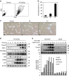
Figure 1
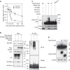
Figure 1. Identification of Fbxo22 as a protein predominantly expressed in larger sized senescent cells and regulation of its expression by p53.
(a) HCA2 cells at 72 h after IR treatment (10 Gy) were sorted according to size and structure (FSC and SSC) and fractionated as P1 and P2 by FACScan. In the absence of IR (Control), the vast majority of cells were fractionated into the P2 fraction. (b) HCA2 cells at 72 h after IR irradiation (10 Gy) were sorted by FSC and SSC into P1 and P2 fractions as shown in Fig. 1a. The sorted cells, as well as HCA2 cells without IR irradiation as a control, were cultured for an additional 3 days, and then subjected to an SA-β-gal assay. (c) Representative images of cells in b are shown. (d) Lysates from HCA2 cells collected at the indicated times after IR (10 Gy) treatment were subjected to immunoblotting using the indicated antibodies. (e) RPE cells expressing Dox-inducible control shRNA (shControl) or p53 shRNA (shp53) were treated with doxycycline (1 μg ml−1). Cell lysates (upper panels) or total RNAs (lower panels) of cells collected at the indicated times after treatment with Nutlin 3a (5 μg ml−1) were subjected to immunoblotting using the indicated antibodies or qPCR analysis, respectively. Data are presented as means ±s.d. of at least three independent experiments.
Figure 2
Figure 2. Fbxo22 controls cell proliferation through regulation of the p53 level.
(a) RPE cells expressing the Dox-inducible shControl or shFbxo22 with or without either Dox-inducible Fbxo22 or shp53 were treated with doxycycline (1 μg ml−1). Lysates of cells collected at the indicated times were subjected to immunoblotting using the indicated antibodies. (b) The relative numbers of cells indicated as in a were determined at the indicated times. (c) RPE cells expressing Dox-inducible wild-type Fbxo22 (Wt), its mutants lacking the F-box domain (ΔF), FIST-C (ΔFIST-C) or FIST-N (ΔFIST-N), as well as control RPE cells (Control), were treated with doxycycline (1 μg ml−1). Lysates of cells collected at the indicated times were subjected to immunoblotting using the indicated antibodies. Because of the antibody epitope, Fbxo22 antibodies failed to recognize ΔFIST-C and ΔFIST-N proteins. (d) The relative numbers of cells as in c were determined at the indicated times. Data are presented as means ±s.d. of at least three independent experiments.
Figure 3
Figure 3. SCFFbxo22 ubiquitylates p53.
(a) RPE cells expressing the Dox-inducible shControl or shFbxo22 in the presence of doxycycline (1 μg ml−1) for 48 h were treated with 50 μg ml−1 cycloheximide (CHX). Lysates of cells collected at the indicated times were subjected to immunoblotting using the indicated antibodies and the relative p53 intensities were determined using Image J. Data are presented as means±s.d. of at least three independent experiments. (b) U2OS cells were transfected with the indicated expression plasmids including SCF components, and were then incubated with MG132 (20 μg ml−1) for 5 h. Total cell lysates were subjected to immunoprecipitation with the indicated antibodies under native conditions. The resultant precipitates and lysates (Input) were subjected to immunoblotting using the indicated HRP-conjugated antibodies. (c) To prevent the detection of ubiquitylation of both E3 ligase itself and p53-associated proteins, the cell lysates were immunoprecipitated with anti-p53 antibody under denaturing conditions. The resultant immunoprecipitates and lysates (Input) were subjected to immunoblotting using the indicated HRP-conjugated antibodies. (d) RPE cells expressing the Dox-inducible full-length Flag-Fbxo22 (Wt) or its mutant lacking the FIST-C domain (FIST-C), as well as control RPE cells were incubated in the presence of doxycycline (1 μg ml−1) for 48 h and then treated with MG132 (10 μg ml−1) for 6 h. Cell lysates were subjected to immunoblotting using the indicated antibodies.
Figure 4
Figure 4. p53 interacts with Fbxo22 in a phosphorylation-independent manner.
(a) RPE cells expressing Dox-inducible Flag-Fbxo22 were incubated for 48 h in the presence or absence of doxycycline (1 μg ml−1) and were then treated with MG132 (10 μg ml−1) for 2 h. The cell lysates were immunoprecipitated using anti-Flag M2 affinity gel. The resultant immunoprecipitates and lysates (Input) were subjected to immunoblotting using anti-Flag or anti-p53 antibodies. (b) RPE cells were treated with MG132 (10 μg ml−1) for 2 h and the lysates were subjected to immunoprecipitation using the anti-p53 antibody or a control IgG. The resultant immunoprecipitates and lysates (Input) were subjected to immunoblotting using the indicated antibodies. (c) Phosphorylation of p53 did not affect its interaction with Fbxo22. RPE cells expressing Dox-inducible Flag-Fbxo22 were incubated for 48 h in the presence or absence of doxycycline (1 μg ml−1) and were then treated with or without MG132 (10 μg ml−1) for 2 h and/or with IR (6 Gy). The lysates were immunoprecipitated using anti-Flag M2 affinity gel. After immunoprecipitation, some samples were treated with protein phosphatase (PP) for 1 h. The resultant immunoprecipitates and lysates (Input) were subjected to immunoblotting using the indicated antibodies. The asterisk indicates an input sample before PP treatment. (d) Interaction between Fbxo22 and p53 was suppressed by treatment with TSA. RPE cells expressing Dox-inducible Flag-Fbxo22 were incubated for 48 h in the presence or absence of doxycycline (1 μg ml−1) and were then treated with or without TSA (1 mM) for 12 h and/or MG132 (10 μg ml−1) for 2 h. The cell lysates were immunoprecipitated using anti-Flag M2 affinity gel. The resultant immunoprecipitates as well as lysates (Input) were subjected to immunoblotting using the indicated antibodies.
Figure 5
Figure 5. Acetylation of lysines at the CTD of p53 suppresses the interaction with Fbxo22.
(a) p53−/− HCT116 cells expressing Dox-inducible Flag-Fbxo22 together with wild-type EGFP-p53 (Wt) or a mutant p53 lacking the C-terminal 30 amino acids (Δ30) were incubated for 48 h in the presence or absence of doxycycline (1 μg ml−1) and were then treated with MG132 (10 μg ml−1) for 2 h. The lysates were immunoprecipitated using anti-Flag M2 affinity gel. The resultant immunoprecipitates and lysates (Input) were subjected immunoblotting using the indicated antibodies. (b) p53−/− HCT116 cells expressing Dox-inducible Flag-Fbxo22 together with the wild-type or the indicated mutants of EGFP-p53 were incubated for 48 h in the presence or absence of doxycycline (1 μg ml−1) and were then treated with MG132 (10 μg ml−1) for 2 h. The lysates were immunoprecipitated using anti-Flag M2 affinity gel. The resultant immunoprecipitates and lysates (Input) were subjected to immunoblotting using the indicated antibodies. (c) RPE cells expressing the Dox-inducible shControl or shFbxo22 were incubated for 48 h in the presence of doxycycline (1 μg ml−1) and were then treated with MG132 (10 μg ml−1) for 2 h. The lysates were immunoprecipitated using a control IgG, methyl (K370me2)- or acetyl (K370Ac)-specific anti-p53, or anti-p53 antibodies. The resultant immunoprecipitates and lysates (Input) were subjected to immunoblotting using the indicated antibodies.
Figure 6
Figure 6. SCFFbxo22 forms a ternary complex with p53 and KDM4A that targets methylated p53 for degradation.
(a) Schematic representation of the domain structure of Fbxo22. (b) RPE cells expressing Dox-inducible wild-type Flag-Fbxo22 (Wt), and its mutants lacking FIST-N (ΔFIST-N) or FIST-C (ΔFIST-C) were incubated for 48 h in the presence or absence of doxycycline (1 μg ml−1) and were then treated with MG132 (10 μg ml−1) for 2 h. The lysates were immunoprecipitated using anti-Flag M2 affinity gel. The resultant immunoprecipitates and lysates (Input) were subjected to immunoblotting using the indicated antibodies. (c) RPE cells expressing Dox-inducible Flag-Fbxo22 were incubated for 48 h in the presence or absence of doxycycline (1 μg ml−1) and were then treated with MG132 (10 μg ml−1) for 2 h. The lysates were subjected to sequential immunoprecipitation using anti-Flag M2 affinity gel and anti-KDM4A antibodies. The resultant immnoprecipitates and lysates were subjected to immunoblotting using the indicated antibodies. (d) RPE cells expressing the Dox-inducible shControl or shFbxo22 together with wild-type KDM4A (Wt) or its catalytically inactive mutant (H188A) were incubated for 48 h in the presence of doxycycline (1 μg ml−1) and were then treated with MG132 (10 μg ml−1) for 2 h. The lysates were subjected to immunoblotting using the indicated antibodies. (e) RPE cells expressing the Dox-inducible shControl, shKDM4A or shKDM4A+shp53 were treated with doxycycline (1 μg ml−1) and their relative numbers were determined at the indicated times. Data are presented as means±s.d. of at least three independent experiments. (f) RPE cells expressing the Dox-inducible shControl or shFbxo22, together with wild-type Flag-PHF20 (Wt) or its WD domain mutant (W97A/Y103A) were incubated for 48 h in the presence of doxycycline (1 μg ml−1) and were then treated with MG132 (10 μg ml−1) for 2 h. The cell lysates were immunoprecipitated using anti-Flag M2 affinity gel. The resultant immunoprecipitates and lysates (Input) were subjected to immunoblotting using the indicated antibodies. (g) Schematic representation of CTD modifications that protect p53 from SCFFbxo22-targeted degradation.
Figure 7
Figure 7. SCFFbxo22-KDM4A-dependent degradation of p53 is essential for induction of p16 and SASP in senescent cells.
(a) Experimental outline of Fbxo22 or KDM4A depletion in senescent cells. (b) HCA2 cells expressing the Dox-inducible shControl or shFbxo22 treated as in a were subjected to immunoblotting using the indicated antibodies. (c) Total RNAs from HCA2 cells as in b were subjected to 7 days of qPCR analysis using the indicated primers. (d) HCA2 cells expressing the Dox-inducible shControl or shKDM4A treated as in a were subjected to immunoblotting using the indicated antibodies. (e) Total RNAs from HCA2 cells as in d were subjected to 7 days of qPCR analysis using the indicated primers. Data are presented as means±s.d. of at least three independent experiments.
Figure 8
Figure 8. CRISPR/CAS9-mediated disruption of mouse Fbxo22.
(a) Representative Fbxo22 wild-type (+/+), heterozygous (+/−) or nullizygous (−/−) mice at 24 weeks of age. (b) Lysates of tissues from the spleen, thymus, kidney, liver, lung and brain of 24-week-old Fbxo22 wild-type (+/+), heterozygous (+/−) or nullizygous (−/−) mice were analysed by immunoblotting using the indicated antibodies. (c) Genotype analysis of Fbxo22 wild-type (+/+), heterozygous (+/−) and nullizygous (−/−) mice by PCR. (d) Primary MEFs from Fbxo22−/− mice showed severe growth retardation. Relative numbers of primary MEFs from Fbxo22+/+, Fbxo22+/− and Fbxo22−/− mice were determined at the indicated times. Data are presented as means±s.d. of at least three independent experiments. (e) Cell cycle distribution of primary MEFs as in d was determined at 4 days by FACScan. (f) Primary MEFs from Fbxo22−/− mice showed marked accumulations of p53, p21 and Mdm2. Primary MEFs from mice with the indicated genotypes were collected at the indicated times and the lysates were subjected to immunoblotting as shown. (g) Primary MEFs with the indicated genotypes were treated with MG132 (10 μg ml−1) for 2 h. The lysates were immunoprecipitated using a control IgG, methyl (K370me2)-specific anti-p53 or anti-p53 antibodies. The resultant immunoprecipitates and lysates (Input) were subjected to immunoblotting using the indicated antibodies.

Similar articles

Cited by

References

    1. Smith J. R. & Pereira-Smith O. M. Replicative senescence: implications for in vivo aging and tumor suppression. Science 273, 63–67 (1996). - PubMed
    1. Blackburn E. H. Switching and signaling at the telomere. Cell 106, 661–673 (2001). - PubMed
    1. Shay J. W. & Wright W. E. Aging. When do telomeres matter? Science 291, 839–840 (2001). - PubMed
    1. Kuilman T., Michaloglou C., Mooi W. J. & Peeper D. S. The essence of senescence. Genes Dev. 24, 2463–2479 (2010). - PMC - PubMed
    1. Johmura Y. et al.. Necessary and sufficient role for a mitosis skip in senescence induction. Mol. Cell 55, 73–84 (2014). - PubMed

MeSH terms