. 2016 Apr;36(4):663-672.
doi: 10.1161/ATVBAHA.116.307126. Epub 2016 Feb 11.

Profound Actions of an Agonist of Growth Hormone-Releasing Hormone on Angiogenic Therapy by Mesenchymal Stem Cells

Profound Actions of an Agonist of Growth Hormone-Releasing Hormone on Angiogenic Therapy by Mesenchymal Stem Cells

Qunchao Ma et al. Arterioscler Thromb Vasc Biol. 2016 Apr.

Abstract

Objective: The efficiency of cell therapy is limited by poor cell survival and engraftment. Here, we studied the effect of the growth hormone-releasing hormone agonist, JI-34, on mesenchymal stem cell (MSC) survival and angiogenic therapy in a mouse model of critical limb ischemia.

Approach and results: Mouse bone marrow-derived MSCs were incubated with or without 10(-8) mol/L JI-34 for 24 hours. MSCs were then exposed to hypoxia and serum deprivation to detect the effect of preconditioning on cell apoptosis, migration, and tube formation. For in vivo tests, critical limb ischemia was induced by femoral artery ligation. After surgery, mice received 50 μL phosphate-buffered saline or with 1×10(6) MSCs or with 1×10(6) JI-34-reconditioned MSCs. Treatment of MSCs with JI-34 improved MSC viability and mobility and markedly enhanced their capability to promote endothelial tube formation in vitro. These effects were paralleled by an increased phosphorylation and nuclear translocation of signal transducer and activator of transcription 3. In vivo, JI-34 pretreatment enhanced the engraftment of MSCs into ischemic hindlimb muscles and augmented reperfusion and limb salvage compared with untreated MSCs. Significantly more vasculature and proliferating CD31(+) and CD34(+) cells were detected in ischemic muscles that received MSCs treated with JI-34.

Conclusions: Our studies demonstrate a novel role for JI-34 to markedly improve therapeutic angiogenesis in hindlimb ischemia by increasing the viability and mobility of MSCs. These findings support additional studies to explore the full potential of growth hormone-releasing hormone agonists to augment cell therapy in the management of ischemia.

Keywords: angiogenesis effects; growth hormone–releasing hormone; mesenchymal stromal cells.


Figures

Figure 1
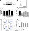
Figure 1. In vitro effects of GHRH agonist, JI-34, on MSC
Expression of GHRH-R on MSC was detected by (A) Western blot and (B) flow cytometry. Grey line is iso-control; black line is GHRH-R. (C) MSCs were treated with JI-34 at different concentrations for 24 hrs, then the medium was replaced with fresh JI-34 free medium. Proliferation of MSCs was evaluated by CCK-8 assay after they were cultured 24 hrs in JI-34 free medium. The maximal effect of GHRH-A was observed at 10 nM (n = 3). *, P < 0.05 vs. 0 nM. (D) The viability of MSC after pretreatment with JI-34 at different concentrations was examined by CCK8 assay after culture under serum deprivation and hypoxic conditions for 48h. (Gray column indicates hypoxia with serum deprivation condition), *, P < 0.05 vs. 0 nM. (E) Flow cytometric analysis of MSC apoptosis. MSC were pretreated with JI-34 (JI) or GHRH antagonist, MIA602 (MIA), for 24 hrs, and then cultured under hypoxia and with serum deprivation medium for 48 hrs. Apoptotic cells (Annexin V+/PI and Annexin V+/PI+ cells) were detected by flow cytometric analysis. (F) Quantifications of apoptotic MSC in E (n = 5-6 in each group). * P < 0.05 vs. 0 nM.
Figure 2
Figure 2. Activation of STAT3 in MSC by JI-34 pretreatment
(A) Western blot analysis of STAT3 activation in lysates from MSC. Dynamic changes of STAT3 phosphorylation were observed at different time points. (B) Expression of phosphorylated STAT3 was quantified as a ratio of phosphorylated STAT3 over total STAT3 by integrated optical density measurement (n = 3). * P < 0.05 vs. control. (C) Immunofluorescence staining for subcellular localization of STAT3 (green) in MSC cultured without additional agent (Control), or with JI-34, or JI-34 + MIA-602, or MIA-602. Scale bars: 50 μm. (D) Quantification of nuclear-localized STAT3 in C by a ratio of fluorescence in nucleus to cytoplasm (n = 3 in each group). * P < 0.05 vs. control.
Figure 3
Figure 3. JI-34 pretreatment enhanced MSC migration and pro-angiogenic effect
(A) Representative images of migration of MSC in a transwell assay. Scale bars: 200 μm. (B) Quantification of migration of MSC. Cells that migrated to the lower chamber were counted (n = 4 in each group). *, P < 0.05 vs. others. (C) Representative images showing tube formation of HUVEC on Matrigel cultured with conditioned media from the specified MSC. Scale bars: 100 μm. (D) Quantification of tube formation in C by measuring branch lengths of formed tube. Only length > 200 μm was counted. n = 5, * P < 0.05 vs. others. (E) Western blot and quantification of pro-angiogenic cytokines expressions in MSCs treated with JI-34 or MIA-602 for 24 hrs. (F) Real time PCR was performed to detect the effect of JI-34 on mRNA expression for VEGF and SDF-1 within MSC. (n = 4) *, P < 0.05 vs. Control.
Figure 4
Figure 4. MSC retention, blood reperfusion, and limb salvage
(A) Donor MSC derived from male mice were injected intramuscularly into female mice. The expression of sry gene in ischemic muscle 3 and 14 days after injection was determined by real-time PCR (n = 3 in each group). *, P < 0.05 vs. MSC group. (B) The blood flow of the lower limbs was quantitatively analyzed as the ratio of ischemic (right) side to nonischemic (left) side (n = 9-15 in each group). *, P < 0.05 vs. PBS; #, P < 0.05 vs. MSC. (C) Representative LDPI images show dynamic changes in blood perfusion ischemic limb at indicated time points. Different colors represent the changes in the perfusion. (D) Representative photographs of hindlimbs from PBS, MSC, or preconditioned MSC treated animals at day 21. (E) Physiological status of ischemic hindlimbs 21 days after transplantation. n = 10 for Sham, 11 for PBS, 13 for MSCs, and 12 for MSC-JI.
Figure 5
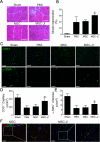
Figure 5. Transplantation of pretreated MSC promotes angiogenesis and muscle regeneration in vivo
(A) Representative hematoxylin and eosin stained sections of ischemic muscles from each group at 21 day; Scale bar: 50 μm, myocytes with centralized nuclei were considered as regenerating myofibers. (B) Quantification of regenerating myofibers by counting the myocytes with centralized nuclei as a percentage of total myocytes in a field (n = 5). *, P < 0.05 vs. PBS and MSC. (C) Immunofluorescent staining of CD31 and α-SMA in cryosections of muscles obtained from mice at day 21 after surgery. Endothelial cells were stained with CD31 and smooth muscle cells were stained with α-SMA. Scale bars: 100μm. (D and E) Quantification of CD31 positive Endothelial cells and α-SMA positive arteriole density (n = 4 in Sham group; n = 5 in PBS group, and other group n = 6). *, P < 0.05 vs. PBS and MSC. (F) Differentiation of MSC in vivo. MSC or MSC preconditioned with JI-34 were stained with DiI (red) and injected into ischemic muscle. The muscles were harvested 21 days later, and cryo-sections stained with DAPI for nuclei (blue) and antibody against CD31 (Green) for endothelial cells. No co-localized DiI with CD31 staining was observed. Scalar bar: 100 μm.
Figure 6
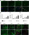
Figure 6. Cell proliferation and CD34+ cell recruitment in ischemic region
Ischemic muscles were harvested 3 or 7 days after surgery. Immunofluorescence staining was performed on the frozen sections of the recovered muscles. (A) Representative sections of ischemic gastrocnemius muscle from day 7 were stained for Ki67 proliferation marker (green), CD31 (red), and DAPI nuclear (blue). A Ki67 and CD31 co-localized cells (pointed by arrows) were demonstrated as the proliferating endothelial cells. Scale bars: 100 μm. (B and C) Quantification of proliferating cells and proliferating endothelial cells, respectively (n = 5 in each group). *, P < 0.05 vs. PBS and MSC. (D) Representative pictures visualized by autofluorescence (green), CD34 (red) and total nuclei (blue) in ischemic gastrocnemius muscle at day 3. (E) Quantification of CD34+ cells (n = 3-4 per group). Scale bars: 100 μm. * P < 0.05 vs. PBS and MSC.
Figure 7
Figure 7. Analysis of proteins related to apoptosis in ischemic muscles
(A) Representative blots of apoptotic related protein expression. (B) Quantification of protein expression levels (n = 3 in each group). *, P < 0.05 vs. PBS group; #, P < 0.05 vs. MSC group.

References

    1. Anversa P, Kajstura J, Rota M, Leri A. Regenerating new heart with stem cells. J Clin Invest. 2013;123:62–70. - PMC - PubMed
    1. Fadini GP, Losordo D, Dimmeler S. Critical reevaluation of endothelial progenitor cell phenotypes for therapeutic and diagnostic use. Circ Res. 2012;110:624–637. - PMC - PubMed
    1. Zhang J, Wilson GF, Soerens AG, Koonce CH, Yu J, Palecek SP, Thomson JA, Kamp TJ. Functional cardiomyocytes derived from human induced pluripotent stem cells. Circ Res. 2009;104:e30–41. - PMC - PubMed
    1. Foldes G, Mioulane M, Wright JS, Liu AQ, Novak P, Merkely B, Gorelik J, Schneider MD, Ali NN, Harding SE. Modulation of human embryonic stem cell-derived cardiomyocyte growth: A testbed for studying human cardiac hypertrophy? J Mol Cell Cardiol. 2011;50:367–376. - PMC - PubMed
    1. Traverse JH, Henry TD, Ellis SG, et al. Effect of intracoronary delivery of autologous bone marrow mononuclear cells 2 to 3 weeks following acute myocardial infarction on left ventricular function: The latetime randomized trial. JAMA. 2011;306:2110–2119. - PMC - PubMed

Publication types

MeSH terms

Substances