

miR-139 Functions as An Antioncomir to Repress Glioma Progression Through Targeting IGF-1 R, AMY-1, and PGC-1β
- PMID: 26868851
- PMCID: PMC5616056
- DOI: 10.1177/1533034616630866
miR-139 Functions as An Antioncomir to Repress Glioma Progression Through Targeting IGF-1 R, AMY-1, and PGC-1β
Abstract
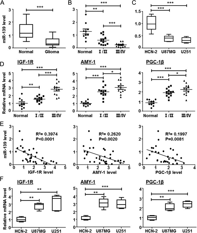
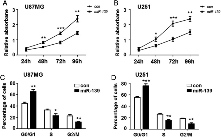
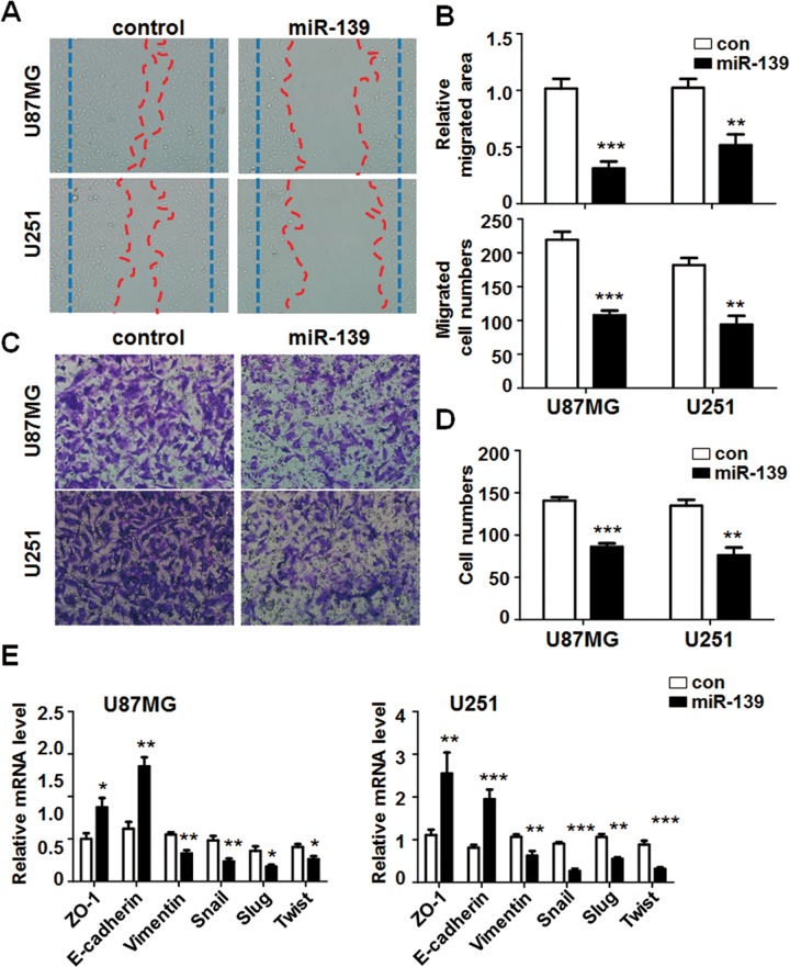
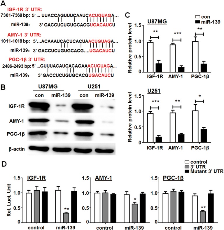
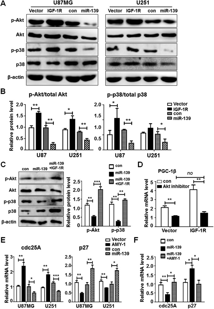
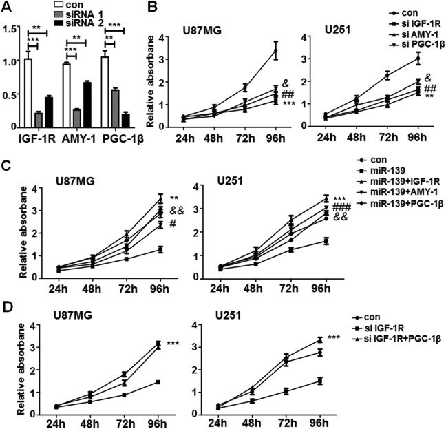
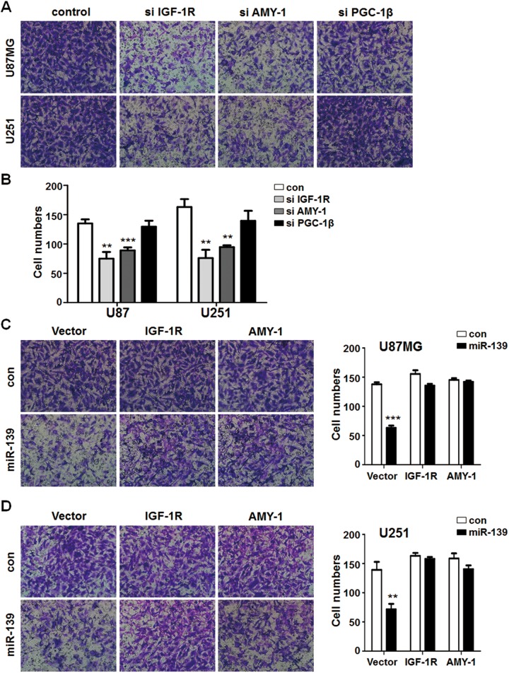
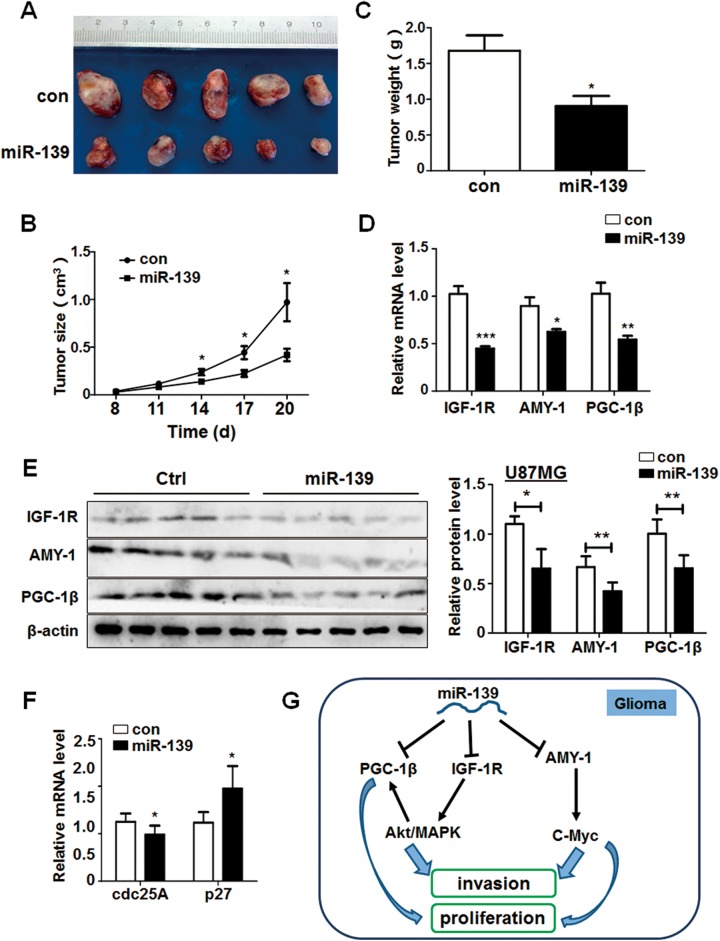
Gliomas are the most common primary malignant brain tumor with poor prognosis, characterized by a highly heterogeneous cell population, extensive proliferation, and migration. A lot of molecular mechanisms regulate gliomas development and invasion, including abnormal expression of oncogenes and variation of epigenetic modification. MicroRNAs could affect cell growth and functions. Several reports have demonstrated that miR-139 plays multifunctions in kinds of solid tumors through different pathways. However, the antitumor mechanisms of this miR-139 are not unveiled in detail. In this study, we not only validated the low expression level of miR-139 in glioma tissues and cell lines but also detected the effect of miR-139 on modulating gliomas proliferation and invasion both in vitro and in vivo. We identified insulin-like growth factor 1 receptor, associate of Myc 1, and peroxisome proliferator-activated receptor γ coactivator 1β as direct targets of miR-139 and the levels of them were all inversely correlated with miR-139 in gliomas. Insulin like growth factor 1 receptor promoted gliomas invasion through Akt signaling and increased proliferation in the peroxisome proliferator-activated receptor γ coactivator 1β-dependent way. Associate of Myc 1 also facilitated gliomas progression by activating c-Myc pathway. Overexpression of the target genes could retrieve the antitumor function of miR-139, respectively, in different degrees. The nude mice transplantation tumor experiment displayed that glioma cells stably expressed miR-139 growth much slower in vivo than the negative control cells. Taken together, these findings suggested miR-139 acted as a favorable factor against gliomas progression and uncovered a novel regulatory mechanism, which may provide a new evidenced prognostic marker and therapeutic target for gliomas.
Keywords: AMY-1; IGF-1 R; PGC-1β; glioma; miR-139; tumor progression.
Conflict of interest statement
Figures
Similar articles
-
MicroRNA-7 directly targets insulin-like growth factor 1 receptor to inhibit cellular growth and glucose metabolism in gliomas.Diagn Pathol. 2014 Nov 14;9:211. doi: 10.1186/s13000-014-0211-y. Diagn Pathol. 2014. PMID: 25394492 Free PMC article.
-
The role of transcriptional coactivator TAZ in gliomas.Oncotarget. 2016 Dec 13;7(50):82686-82699. doi: 10.18632/oncotarget.12625. Oncotarget. 2016. PMID: 27764783 Free PMC article.
-
MicroRNA-144 represses gliomas progression and elevates susceptibility to Temozolomide by targeting CAV2 and FGF7.Sci Rep. 2020 Mar 5;10(1):4155. doi: 10.1038/s41598-020-60218-9. Sci Rep. 2020. PMID: 32139705 Free PMC article.
-
MicroRNAs involved in the EGFR/PTEN/AKT pathway in gliomas.J Neurooncol. 2012 Jan;106(2):217-24. doi: 10.1007/s11060-011-0679-1. Epub 2011 Aug 13. J Neurooncol. 2012. PMID: 21842313 Review.
-
The emerging role of miR-19 in glioma.J Cell Mol Med. 2018 Oct;22(10):4611-4616. doi: 10.1111/jcmm.13788. Epub 2018 Aug 2. J Cell Mol Med. 2018. PMID: 30073755 Free PMC article. Review.
Cited by
-
miR-139/PDE2A-Notch1 feedback circuit represses stemness of gliomas by inhibiting Wnt/β-catenin signaling.Int J Biol Sci. 2021 Aug 12;17(13):3508-3521. doi: 10.7150/ijbs.62858. eCollection 2021. Int J Biol Sci. 2021. PMID: 34512162 Free PMC article.
-
A panel of eight microRNAs is a good predictive parameter for triple-negative breast cancer relapse.Theranostics. 2020 Jul 9;10(19):8771-8789. doi: 10.7150/thno.46142. eCollection 2020. Theranostics. 2020. PMID: 32754277 Free PMC article.
-
Targeting the Notch1 oncogene by miR-139-5p inhibits glioma metastasis and epithelial-mesenchymal transition (EMT).BMC Neurol. 2018 Aug 31;18(1):133. doi: 10.1186/s12883-018-1139-8. BMC Neurol. 2018. PMID: 30170559 Free PMC article.
-
miR‑497/MIR497HG inhibits glioma cell proliferation by targeting CCNE1 and the miR‑588/TUSC1 axis.Oncol Rep. 2021 Dec;46(6):255. doi: 10.3892/or.2021.8206. Epub 2021 Oct 19. Oncol Rep. 2021. PMID: 34664678 Free PMC article.
-
MicroRNA-139 targets fibronectin 1 to inhibit papillary thyroid carcinoma progression.Oncol Lett. 2017 Dec;14(6):7799-7806. doi: 10.3892/ol.2017.7201. Epub 2017 Oct 17. Oncol Lett. 2017. Retraction in: Oncol Lett. 2022 May 31;24(1):237. doi: 10.3892/ol.2022.13357. PMID: 29250177 Free PMC article. Retracted.
References
-
- Wen PY, Kesari S. Malignant gliomas in adults. N Engl J Med. 2008;359(5):492–507. - PubMed
-
- Stupp R, Mason WP, van den Bent MJ, et al. Radiotherapy plus concomitant and adjuvant temozolomide for glioblastoma. N Engl J Med. 2005;352(10):987–996. - PubMed
-
- Butowski NA, Sneed PK, Chang SM. Diagnosis and treatment of recurrent high-grade astrocytoma. J Clin Oncol. 2006;24(8):1273–1280. - PubMed
MeSH terms
Substances
LinkOut - more resources
Full Text Sources
Other Literature Sources
Medical
Miscellaneous
