. 2016 Jul;29(7):318-33.
doi: 10.1002/jmr.2533. Epub 2016 Feb 12.

Grb7 and Hax1 may colocalize partially to mitochondria in EGF-treated SKBR3 cells and their interaction can affect Caspase3 cleavage of Hax1

Affiliations

Grb7 and Hax1 may colocalize partially to mitochondria in EGF-treated SKBR3 cells and their interaction can affect Caspase3 cleavage of Hax1

Lei Qian et al. J Mol Recognit. 2016 Jul.

Abstract

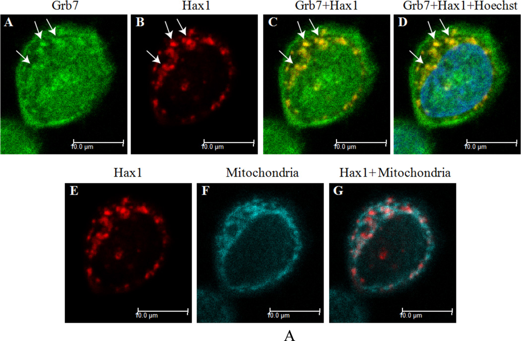
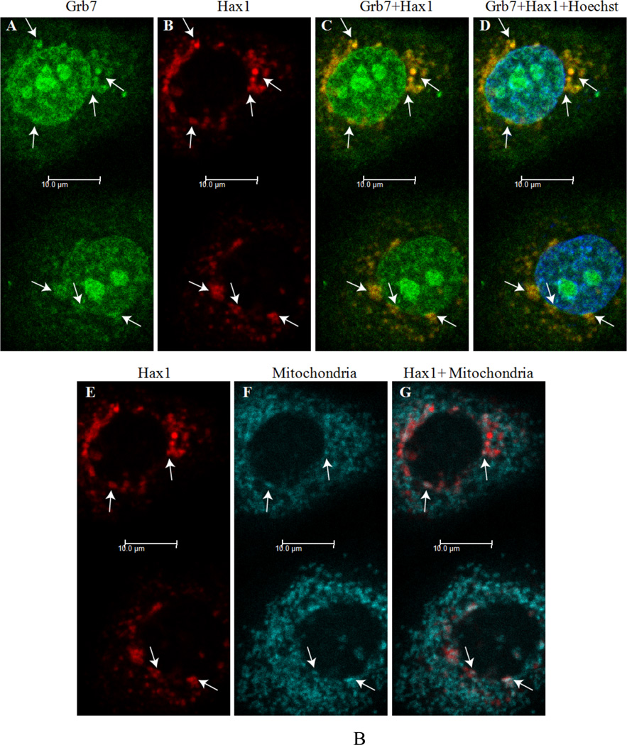
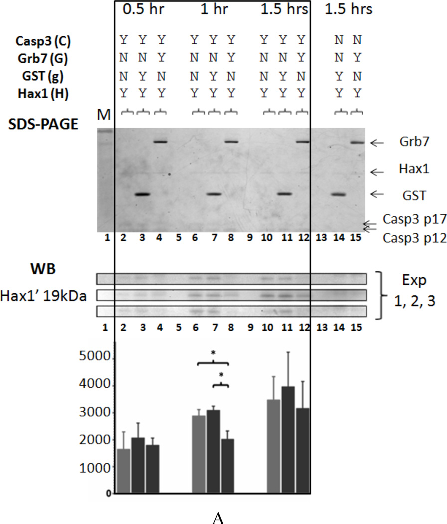
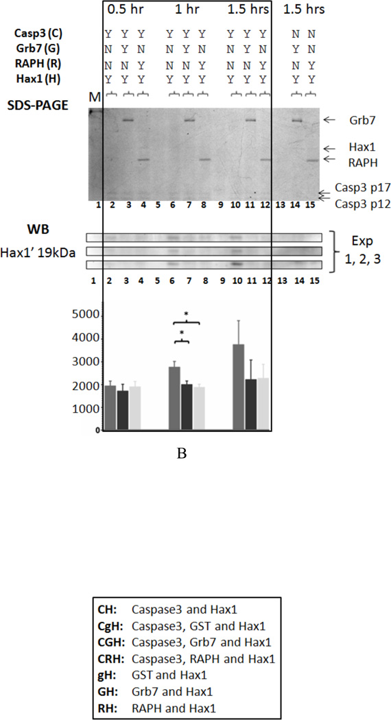
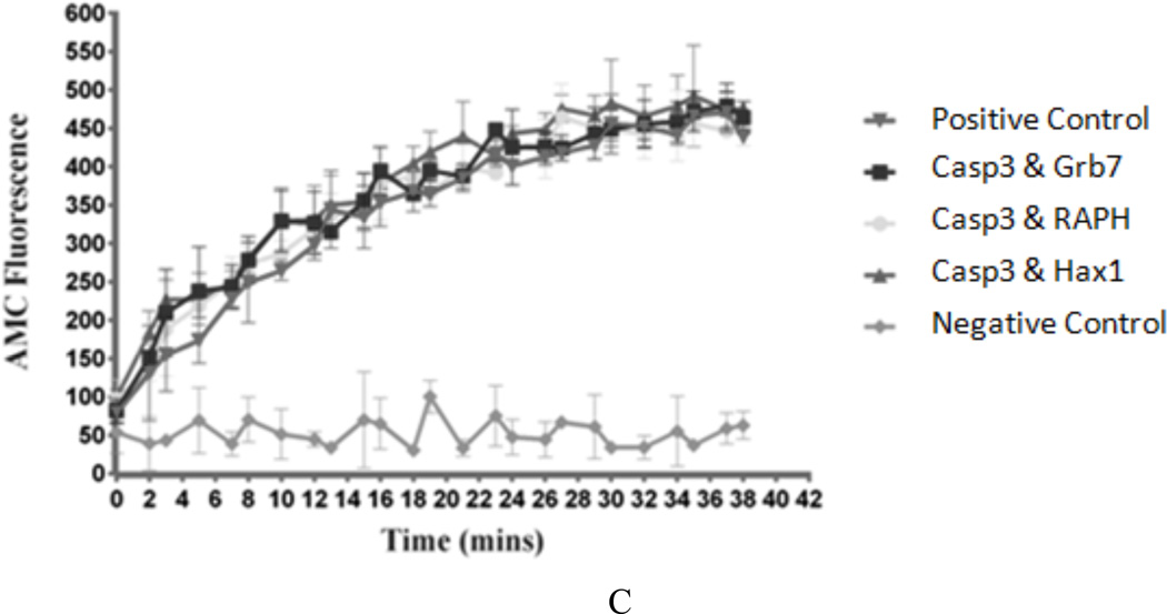
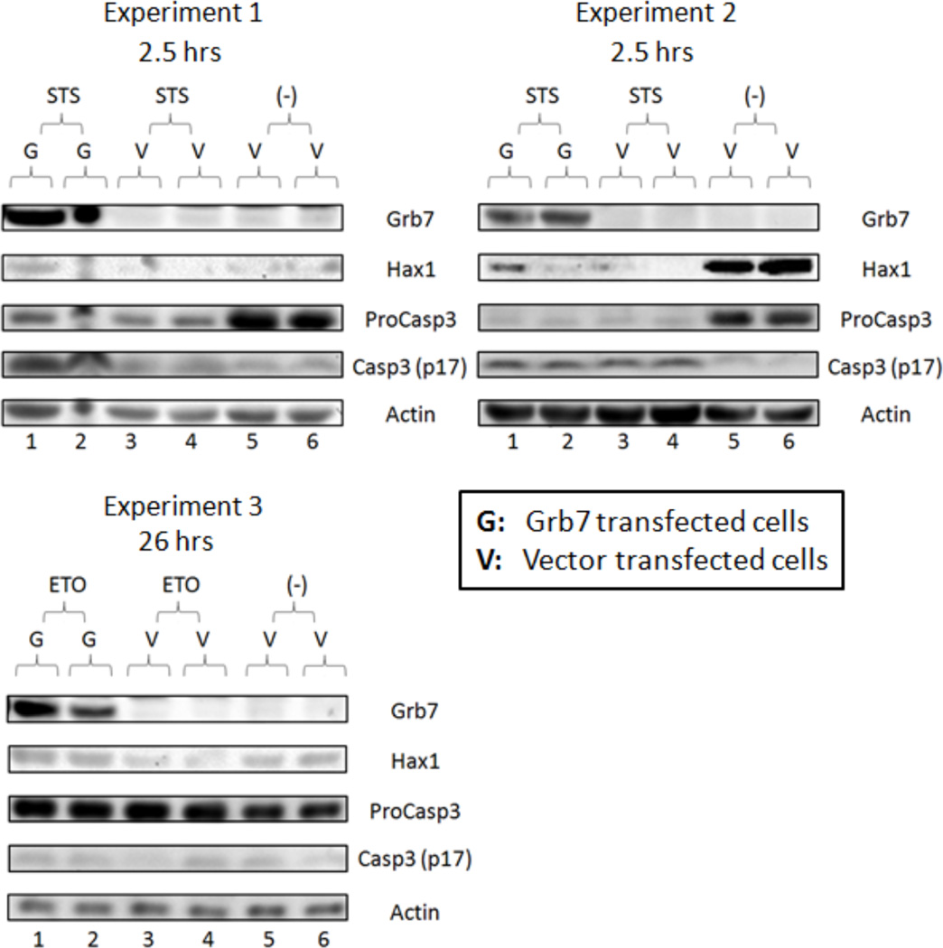
Growth factor receptor bound protein 7 (Grb7) is a signal-transducing adaptor protein that mediates specific protein-protein interactions in multiple signaling pathways. Grb7, with Grb10 and Grb14, is members of the Grb7 protein family. The topology of the Grb7 family members contains several protein-binding domains that facilitate the formation of protein complexes, and high signal transduction efficiency. Grb7 has been found overexpressed in several types of cancers and cancer cell lines and is presumed involved in cancer progression through promotion of cell proliferation and migration via interactions with the erythroblastosis oncogene B 2 (human epidermal growth factor receptor 2) receptor, focal adhesion kinase, Ras-GTPases, and other signaling partners. We previously reported Grb7 binds to Hax1 (HS1 associated protein X1) isoform 1, an anti-apoptotic protein also involved in cell proliferation and calcium homeostasis. In this study, we confirm that the in vitro Grb7/Hax1 interaction is exclusive to these two proteins and their interaction does not depend on Grb7 dimerization state. In addition, we report Grb7 and Hax1 isoform 1 may colocalize partially to mitochondria in epidermal growth factor-treated SKBR3 cells and growth conditions can affect this colocalization. Moreover, Grb7 can affect Caspase3 cleavage of Hax1 isoform 1 in vitro, and Grb7 expression may slow Caspase3 cleavage of Hax1 isoform 1 in apoptotic HeLa cells. Finally, Grb7 is shown to increase cell viability in apoptotic HeLa cells in a time-dependent manner. Taken together, these discoveries provide clues for the role of a Grb7/Hax1 protein interaction in apoptosis pathways involving Hax1. Copyright © 2016 John Wiley & Sons, Ltd.

Keywords: Caspase3; Grb7; Hax1; SKBR3 cells; apoptosis; mitochondria; signal-transducing adaptor protein.


Figures

Figure 1
Figure 1. A) Top: Domain topology of the human Grb7 protein isoform 1
The approximate amino acid residue numbers defining each domain are indicated by numbers. Bottom: Domain topology of the human Hax1 protein isoform 1 The approximate amino acid residue numbers defining each domain or motif are indicated by numbers. B) Western Blot results for the in vitro binding assay of purified SUMO-Grb7-RAPH domains and purified Hax1 Lane 1: Last wash sample from the negative control. Lane 2: Last wash sample from the in vitro binding assay of SUMO-RAPH and Hax1. Lane 3: Blank. Lane 4–6: Elution samples (three elutions) from the in vitro binding assay of SUMO-RAPH and Hax1. Lane 7–9: Elution samples (three elutions) from the negative control. C) Western Blot results for the in vitro binding assay of purified Grb7 or Grb7 (F511R) and purified Hax1 Lane 1: Last wash sample from the in vitro binding assay of Grb7 and Hax1. Lane 2: Last wash sample from the in vitro binding assay of Grb7 (F511R) and Hax1. Lane 3: Last wash sample from the negative control. Lane 4–5: Elution samples (two elutions) from the in vitro binding assay of Grb7 and Hax1. Lane 6: Blank. Lane 7–8: Elution samples (two elutions) from the in vitro binding assay of Grb7 (F511R) and Hax1. Lane 9: Blank. Lane 10–11: Elution samples (two elutions) from the negative control.
Figure 2
Figure 2. A) Grb7 and Hax1 isoform 1 colocalize in SKBR3 cells stained with mitochondria marker
Two days post-transfection cells were stained with mitochondria marker. Colocalization sites of Grb7 and Hax1 are indicated by arrows and the overlapping yellow color in Panels C and D. Scale bar = 10 µm. B) Grb7 and Hax1 isoform 1 colocalize in EGF treated SKBR3 cells stained with mitochondria marker Two days post-transfection cells were serum starved, treated with EGF, and stained with mitochondria marker. Colocalization sites of Grb7 and Hax1 are indicated by arrows and the overlapping yellow color in Panels C and D. Possible colocalization sites with mitochondria are shown by arrows in Panel G. Scale bar = 10 µm.
Figure 3
Figure 3. A) Top: Enzyme inhibition model for Caspase3 (enzyme), Hax1 (substrate) and Grb7 (inhibitor)
In the proposed model Grb7 (indicated by the circled “I” letter) acts as a substrate inhibitor as described in the text. Bottom: Model of Grb7 inhibition in Caspase3 cleavage of Hax1 Grb7 as an inhibitor binds to Hax1, affecting Caspase3 cleavage of Hax1. B) Caspase3 cleavage site on Hax1 Caspase3 cleaves between D127 and S128 of Hax1, producing the 14kDa and 19kDa cleavage products discussed in the text.
Figure 4
Figure 4. A) Coomassie Blue staining, Western Blot, and densitometric analysis results for the in vitro Caspase3 cleavage assay I
Lane 1: Protein Marker. Lane 2: CH: Caspase3 and Hax1 at 0.5 hr. Lane 3: CgH: Caspase3, GST and Hax1 at 0.5 hr. Lane 4: CGH: Caspase3, Grb7 and Hax1 at 0.5 hr. Lane 5: Blank. Lane 6: CH at 1 hr. Lane 7: CgH at 1 hr. Lane 8: CGH at 1 hr. Lane 9: Blank. Lane 10: CH at 1.5 hrs. Lane 11: CgH at 1.5 hrs. Lane 12: CGH at 1.5 hrs. Lane 13: Blank. Lane 14: gH: GST and Hax1 at 1.5 hrs. Lane 15: GH: Grb7 and Hax1 at 1.5 hrs. The assay was repeated three times and the amounts of cleavage product Hax1’ (19kDa) were studied by Western Blot and densitometric analysis. Asterisks indicate statistically relevant results (P values < 0.02) B) Coomassie Blue staining, Western Blot and densitometric analysis results for the in vitro Caspase3 cleavage assay II Lane 1: Protein Marker. Lane 2: CH: Caspase3 and Hax1 at 0.5 hr. Lane 3: CGH: Caspase3, Grb7 and Hax1 at 0.5 hr. Lane 4: CRH: Caspase3, RAPH and Hax1 at 0.5 hr. Lane 5: Blank. Lane 6: CH at 1 hr. Lane 7: CGH at 1 hr. Lane 8: CRH at 1 hr. Lane 9: Blank. Lane 10: CH at 1.5 hrs. Lane 11: CGH at 1.5 hrs. Lane 12: CRH at 1.5 hrs. Lane 13: Blank. Lane 14: GH: Grb7 and Hax1 at 1.5 hrs. Lane 15: RH: RAPH and Hax1 at 1.5 hrs. The assay was repeated three times and the cleavage product Hax1’ (19kDa) was analyzed by Western Blot and densitometric analysis. Asterisks indicate statistically relevant results (P values < 0.02). C) in vitro Caspase3 activity assay Five different cases are shown: Ac-DEVD-AMC only (negative control), Caspase3 with Ac-DEVD-AMC (positive control), and Grb7-6xHis, RAPH-6xHis or Hax1-6xHis incubated with Caspase3 and Ac-DEVD-AMC. The fluorescence signal from released AMC was recorded every 1–2 minutes for a total of 40 minutes.
Figure 5
Figure 5. Grb7 may slow cleavage or degradation of Hax1 isoform 1 in apoptotic HeLa cells. Top (Experiments 1 and 2): Western Blot results for two STS induced apoptosis trials in HeLa cells
Lane 1–2: cell lysates from apoptotic Grb7 transfected cells. Lane 3–4: cell lysates from apoptotic vector transfected cells. Lane 5–6: untreated vector transfected cell lysates. Bottom (Experiment 3): Western Blot results for a single ETO induced apoptosis trial in HeLa cells Lane 1–2: cell lysates from apoptotic Grb7 transfected cells. Lane 3–4: cell lysates from apoptotic vector transfected cells. Lane 5–6: untreated vector transfected cell lysates.
Figure 6
Figure 6. Top: Western Blot results for transfected cells prior to STS induced apoptosis
Lane 1–6: Lysates from HeLa cells transfected with different sets of plasmids. V: pcDNA3.1(+) vector, G: pcDNA3.1(+)-Grb7, H: pcDNA3.1(+)-Hax1, C: pcDNA3.1(+)-ProCaspase3. Bottom: CellTiter-Glo luminescent cell viability assay for transfected cells after STS induced apoptosis Viability ratios for all protein expression combinations (indicated in the figure legend) are graphically reported at the 1.5 hrs and 2.5 hrs time points. Asterisks indicate statistically relevant results (P values < 0.02).

Similar articles

Cited by

References

    1. Cavnar PJ, Berthier E, Beebe DJ, Huttenlocher A. Hax1 regulates neutrophil adhesion and motility through RhoA. J Cell Biol. 2011;193(3):465–473. - PMC - PubMed
    1. Chae HJ, Kang JS, Byun JO, Han KS, Kim DU, Oh SM, Kim HM, Chae SW, Kim HR. Molecular mechanism of staurosporine-induced apoptosis in osteoblasts. Pharmacol Res. 2000;42(4):373–381. - PubMed
    1. Chandra D, Tang DG. Mitochondrially localized active caspase-9 and caspase-3 result mostly from translocation from the cytosol and partly from caspase-mediated activation in the organelle. Lack of evidence for Apaf-1-mediated procaspase-9 activation in the mitochondria. J Biol Chem. 2003;278(19):17408–17420. - PubMed
    1. Chao JR, Parganas E, Boyd K, Hong CY, Opferman JT, Ihle JN. Hax1-mediated processing of HtrA2 by Parl allows survival of lymphocytes and neurons. Nature. 2008;452(7183):98–102. - PubMed
    1. Chu PY, Li TK, Ding ST, Lai IR, Shen TL. EGF-induced Grb7 recruits and promotes Ras activity essential for the tumorigenicity of Sk-Br3 breast cancer cells. J Biol Chem. 2010;285(38):29279–29285. - PMC - PubMed