. 2016 Feb 12:6:20879.
doi: 10.1038/srep20879.

Prostaglandin I₂ Attenuates Prostaglandin E₂-Stimulated Expression of Interferon γ in a β-Amyloid Protein- and NF-κB-Dependent Mechanism

Affiliations

Prostaglandin I₂ Attenuates Prostaglandin E₂-Stimulated Expression of Interferon γ in a β-Amyloid Protein- and NF-κB-Dependent Mechanism

Pu Wang et al. Sci Rep. .

Erratum in

Abstract

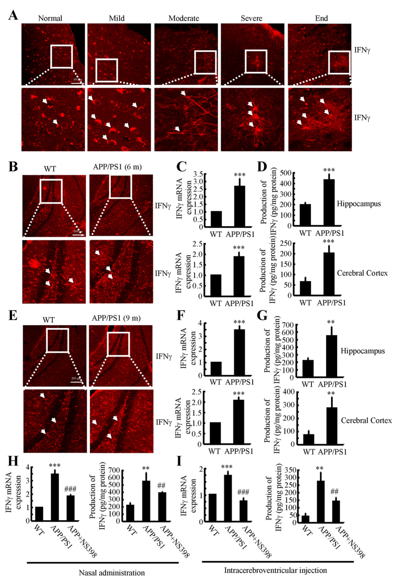
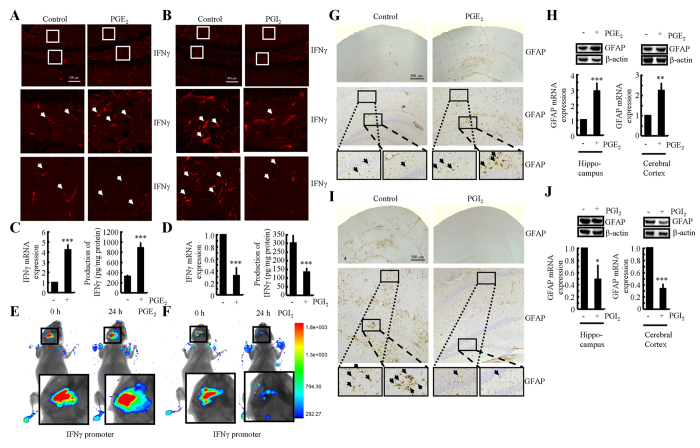
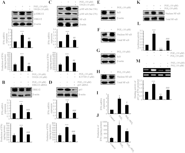
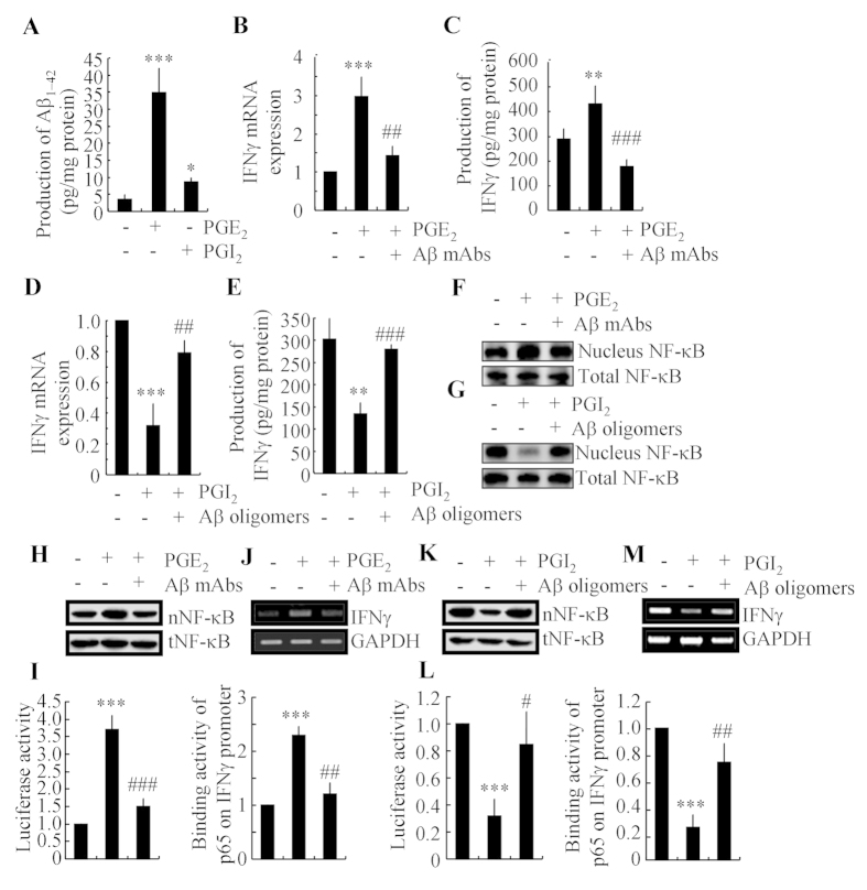
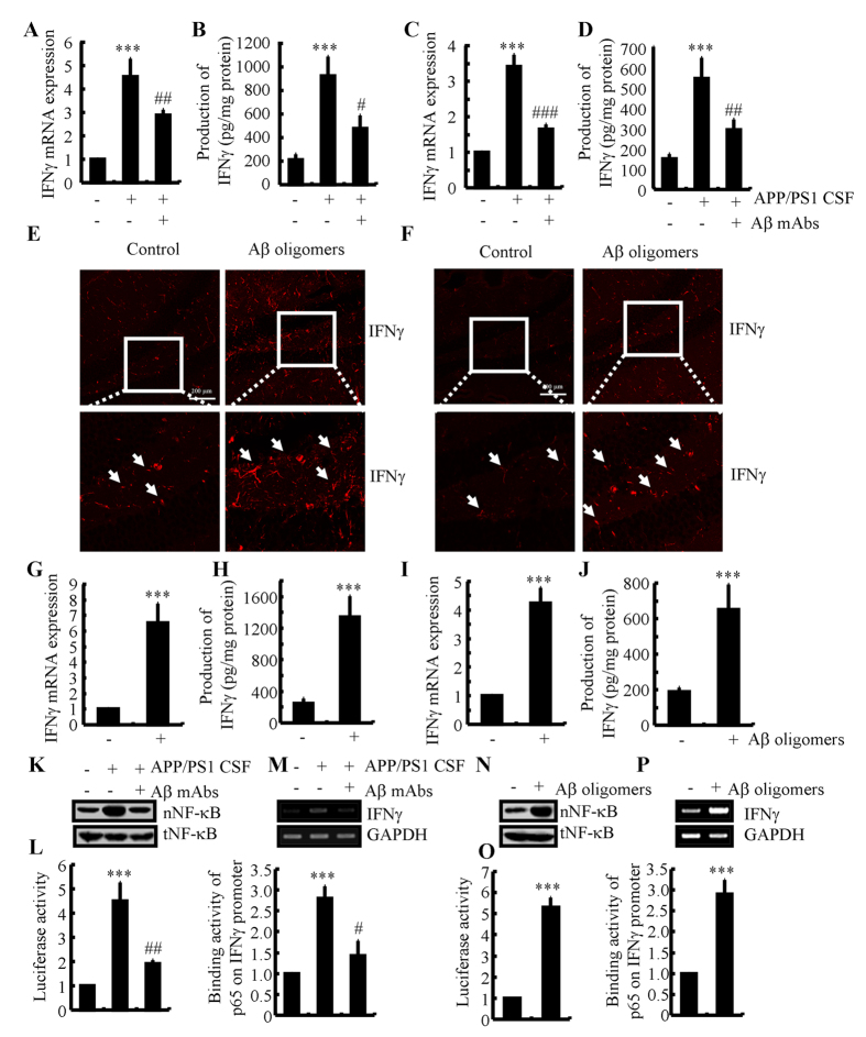
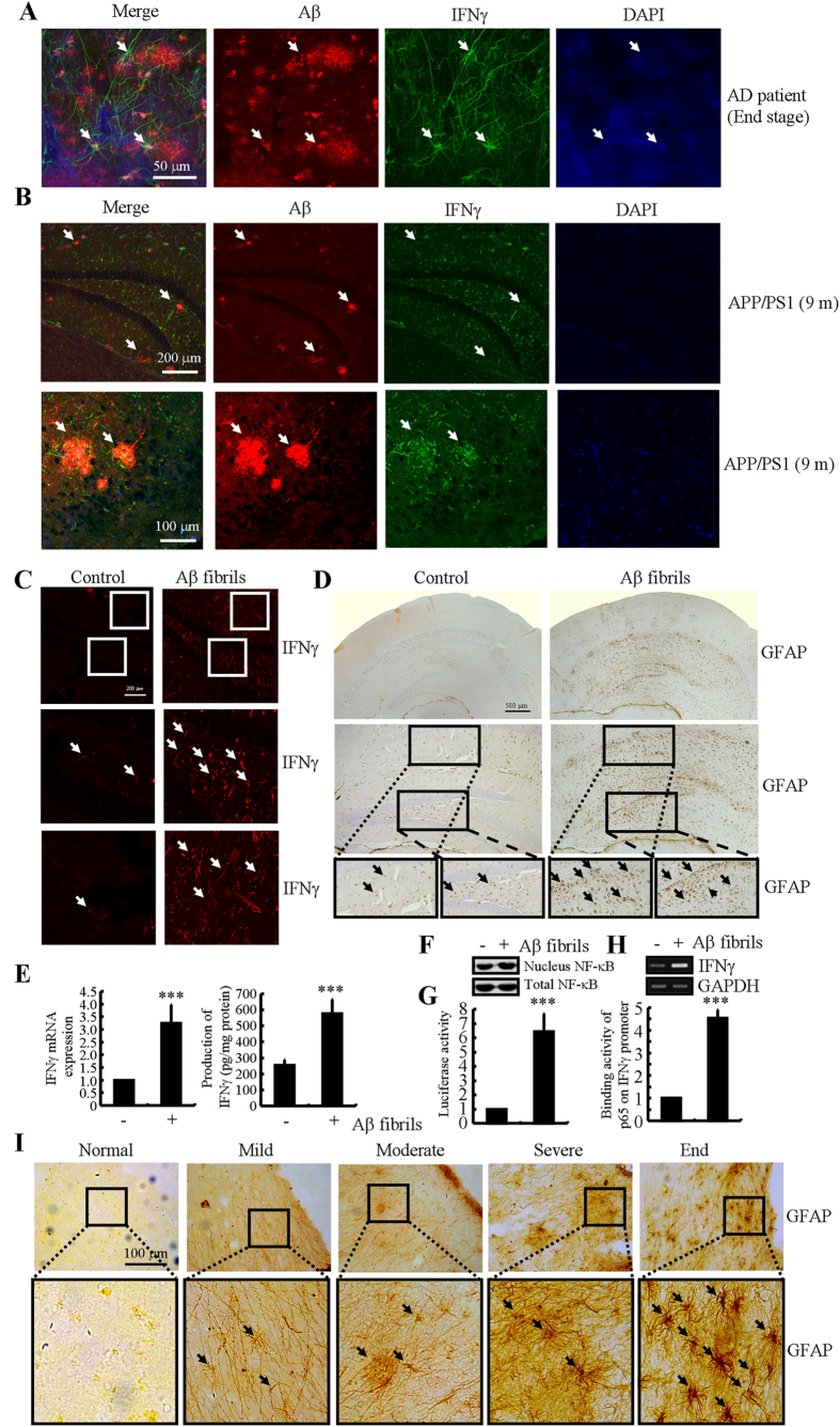
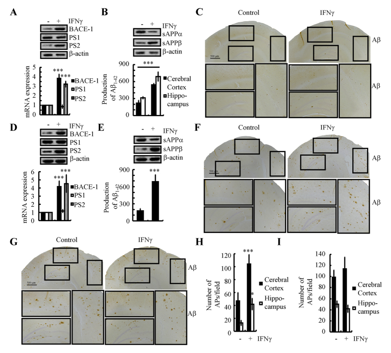
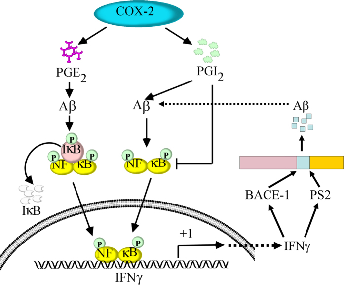
Cyclooxygenase-2 (COX-2) has been recently identified as being involved in the pathogenesis of Alzheimer's disease (AD). However, the role of an important COX-2 metabolic product, prostaglandin (PG) I2, in AD development remains unknown. Using mouse-derived astrocytes as well as APP/PS1 transgenic mice as model systems, we firstly elucidated the mechanisms of interferon γ (IFNγ) regulation by PGE2 and PGI2. Specifically, PGE2 accumulation in astrocytes activated the ERK1/2 and NF-κB signaling pathways by phosphorylation, which resulted in IFNγ expression. In contrast, the administration of PGI2 attenuated the effects of PGE2 on stimulating the production of IFNγ via inhibiting the translocation of NF-κB from the cytosol to the nucleus. Due to these observations, we further studied these prostaglandins and found that both PGE2 and PGI2 increased Aβ1-42 levels. In detail, PGE2 induced IFNγ expression in an Aβ1-42-dependent manner, whereas PGI2-induced Aβ1-42 production did not alleviate cells from IFNγ inhibition by PGI2 treatment. More importantly, our data also revealed that not only Aβ1-42 oligomer but also fibrillar have the ability to induce the expression of IFNγ via stimulation of NF-κB nuclear translocation in astrocytes of APP/PS1 mice. The production of IFNγ finally accelerated the deposition of Aβ1-42 in β-amyloid plaques.


Figures

Figure 1
Figure 1. NS398 treatment decreases the induction of IFNγ in APP/PS1 mice.
(A) The tissue blocks of human brains at different stages of AD were collected by the New York Brain Bank at Columbia University. Free-floating slices (40 μm) were prepared by cryostat. (B–G) The brains of WT or APP/PS1 transgenic mice at 6 or 9 months of age were collected following anesthesia and perfusion. In select experiments, the APP/PS1 transgenic mice at the age of 3 month received NS398 (50 μg/kg/d) intranasally for 6 months before brain harvesting (H). In separate experiments, APP/PS1 mice were injected (i.c.v) with NS398 (2 μg/5 μl) for 24 h (I). The immunoreactivity of IFNγ was determined by immunohistochemistry using an anti-IFNγ antibody (A,B,E). The arrows demonstrated the positive staining of IFNγ. IFNγ protein and mRNA levels were determined by IFNγ enzyme immunoassay kits and qRT-PCR, respectively (C–I). Total amounts of protein and GAPDH served as an internal control. The data represent the means ± S.E. of atleast three independent experiments. *p < 0.05; **p < 0.01 and ***p < 0.001 with respect to WT control. #p < 0.05; ##p < 0.01 and ###p < 0.001 compared to APP/PS1 alone.
Figure 2
Figure 2. Antagonistic effects of PGE2 and PGI2 on regulating the expression of IFNγ in WT or APP/PS1 transgenic mice.
The WT or APP/PS1 C57BL/6 mice at the age of 6 months were injected (i.c.v.) with PGE2 (2 μg/5 μl) or PGI2 (2 μg/5 μl). The brains were then collected and sectioned after 24 h (A,B,G,I). In select experiments, one side of the cerebral ventricle was injected with PGE2 (2 μg/5 μl) or PGI2 (2 μg/5 μl), and the other side of the cerebral ventricle was injected (i.c.v.) with D1A cells, which was pre-transfected with the IFNγ promoter (E,F). The immunoreactivity of IFNγ was determined by immunofluorescence staining using an anti-IFNγ antibody (A,B). Luciferase activities from the different groups of mice were measured using live animal imaging system (E,F). The activities of astrocytes were determined by immunohistochemistry with anti-GFAP (G,I). mRNA and protein levels of IFNγ and GFAP were determined by qRT-PCR, western blot and IFNγ enzyme immunoassay kits, respectively (C,D,H,J). Total amounts of GAPDH, β-actin and protein served as an internal control. The data represent the means ± S.E. of atleast three independent experiments. *p < 0.05; **p < 0.01 and ***p < 0.001 with respect to PBS (−) or vehicle-treated controls.
Figure 3
Figure 3. Critical roles of NF-κB activity in regulating IFNγ expression by PGE2- and PGI2-stimulated D1A cells.
Mouse astrocyte D1A cells were treated with PGE2 (10 μM) in the absence or presence of the ERK1/2 inhibitor U0126 (10 μM) (A,C upper panel), KT5720 (5 μM) (C lower panel) for 24 h before extracting protein or mRNA (A,C,E,F). In select experiments, the cells were transfected with ERK1/2 or p65 siRNA before incubation with PGE2 (10 μM) for 24 h (B,D). In separate experiments, cells were treated with PGI2 (10 μM) for 24 h (G,H). In distinct experiments, the cells were treated with PGE2 (10 μM) in the absence or presence of PGI2 (10 μM) for 24 h (I–M). Total ERK1/2 (A,B), phosphorylated ERK1/2 levels (A), total NF-κB (C,D), phosphorylated NF-κB (C) and total IκB (E,G) were detected by immunoblotting using specific antibodies. Equal lane loading was demonstrated by the similar intensities of total β-actin. The nuclear and total NF-κB levels were determined by western blots (F,H,K). IFNγ protein and mRNA levels were determined by IFNγ enzyme immunoassay kits and qRT-PCR, respectively (A–D,I,J). Total amounts of protein and GAPDH served as an internal control. The luciferase activity of the IFNγ promoter was determined by dual luciferase reporter assay kits (L). The binding activity of NF-κB to the promoter of IFNγ was determined by ChIP assay (M). The data represent the means ± S.E. of atleast three independent experiments. *p < 0.05; **p < 0.01 and ***p < 0.001 with respect to the vehicle-treated or vector-transfected control. #p < 0.05; ##p < 0.01 and ###p < 0.001 compared to PGE2-treated alone.
Figure 4
Figure 4. Aβ1–42 mediated the antagonistic effects of PGE2 and PGI2 on regulating the expression of IFNγ.
D1A cells were treated with PGE2 (10 μM) or PGI2 (10 μM) for 48 h (A). In select experiments, PGE2 (2 μg/5 μl) or PGI2 (2 μg/5 μl) was injected (i.c.v.) into the ventricles of WT C57BL/6 or APP/PS1 mice in the absence or presence of Aβ antibody (1 μg/5 μl) or Aβ1–42 oligomers (1 μg/5 μl) for 24 h (BG). In separate experiments, D1A cells were treated with PGE2 (10 μM) in the absence or presence of Aβ antibody (1 μg/ml) for 24 h (H–J). In distinct experiments, D1A cells were treated with PGI2 (10 μM) in the absence or presence of Aβ1–42 oligomers (1 μM) (KM). The production of Aβ1–42 was determined by Aβ1–42 ELISA kits (A). Total amount of protein served as internal control. IFNγ protein and mRNA levels were determined by IFNγ enzyme immunoassay kits and qRT-PCR, respectively (B–E). Total amounts of protein and GAPDH served as an internal control. The nuclear and total NF-κB levels were determined by western blots (F–K). The luciferase activity of the IFNγ promoter was determined by dual luciferase reporter assay kits (I,L). The binding activity of NF-κB to the promoter of IFNγ was determined by ChIP assay (J,M). The data represent the means ± S.E. of atleast three independent experiments. *p < 0.05; **p < 0.01 and ***p < 0.001 with respect to the vehicle-treated control. #p < 0.05; ##p < 0.01 and ###p < 0.001 compared to PGE2-treated alone.
Figure 5
Figure 5. IFNγ upregulation at the early stage of AD was caused by Aβ oligomers.
Cerebrospinal fluid (CSF) of APP/PS1 mice at 6 months of age was collected and then injected (i.c.v.) into wild type C57BL/6 mice in the absence or presence of Aβ antibody (1 μg/5 μl) for two weeks before sacrifice (A,B). In select experiments, D1A cells were treated with CSF of APP/PS1 mice at 6 months of age (1:1000 dilution) in the absence or presence of Aβ antibody (1 μg/ml) for 24 h (C,D,K–M). In separate experiments, the wild type C57BL/6 mice at the age of 6 months were injected (i.c.v) with Aβ oligomers (2 μg/5 μl) for 24 h (E,G,H). In distinct experiments, the slices of 6-month-old WT mice or D1A cells were cultured in Aβ1–42 oligomers (F,I,J,N–P). The immunoactivity of IFNγ was determined by an immunofluorescence assay (E,F). IFNγ protein and mRNA levels were determined by IFNγ enzyme immunoassay kits and qRT-PCR, respectively (A–D,G–J). Total amounts of protein and GAPDH served as an internal control. The nuclear and total NF-κB levels were determined by western blots (K,N). The luciferase activity of the IFNγ promoter was determined by dual luciferase reporter assay kits (L,O). The binding activity of NF-κB to the promoter of IFNγ was determined by ChIP assay (M,P). The data represent the means ± S.E. of atleast three independent experiments. *p < 0.05; **p < 0.01 and ***p < 0.001 with respect to vehicle-treated controls. #p < 0.05; ##p < 0.01 and ###p < 0.001 compared to APP/PS1 CSF-treated alone.
Figure 6
Figure 6. IFNγ upregulation at the late stage of AD was caused by advanced aggregated Aβ in APs.
(A,I) The tissue blocks of human brains at different stages of AD were collected by the New York Brain Bank at Columbia University. Free-floating slices (40 μm) were prepared by cryostat. (B) The brains of WT or APP/PS1 transgenic mice at 9 months of age were collected after anesthesia and perfusion. In select experiments, brain slices of 6-month-old WT mice were cultured in the absence or presence of Aβ1–42 fibrils for 24 h (C, D). In separate experiments, D1A cells were incubated with Aβ fibers for 24 h (E–H). The slices of mouse brains were double-stained with Aβ (red) or IFNγ (green) antibodies before being observed under confocal microscopy (A,B). The immunoactivity of IFNγ was determined by an immunofluorescence assay (C). The activity of astrocytes was determined by staining with GFAP (D,I). IFNγ protein and mRNA levels were determined by IFNγ enzyme immunoassay kits and qRT-PCR, respectively (E). Total amounts of protein and GAPDH served as an internal control. The nuclear and total NF-κB levels were determined by western blots (F). The luciferase activity of the IFNγ promoter was determined by a dual luciferase reporter assay kits (G). The binding activity of NF-κB to the promoter of IFNγ was determined by a ChIP assay (H). The data represent the means ± S.E. of atleast three independent experiments. *p < 0.05; **p < 0.01 and ***p < 0.001 with respect to vehicle-treated controls.
Figure 7
Figure 7. Nasal administration of IFNγ accelerates Aβ deposition in the brains of APP/PS1 mice by inducing the expression of BACE-1 and PS2.
IFNγ (10 ng/20 μl/d) was nasally administered to 3-months-old WT mice for 7 days (A–C). In select experiments, n2a cells were treated with IFNγ (10 ng/ml) for 24 h before extracting total mRNA and protein (D,E). In separate experiments, 3-months-old APP/PS1 mice was nasally administered with IFNγ (10 ng/20 μl/d) for 3 or 6 months before determining the Aβ deposition in APs (F–I). The protein and mRNA expression of BACE-1, PS1 and PS2 were determined by western blot and qRT-PCR (A,D). Total amounts of β-actin and GAPDH served as an internal control. The production of sAPPα, sAPPβ and Aβ1–42 was determined by western blot and Aβ1–42 enzyme immunoassay kits (B,E). Total amounts of β-actin and protein served as an internal control. The immunoactivity of Aβ was determined by an immunohistochemistry assay (C,F,G). APs/field in cerebral cortex and hippocampus of APP/PS1 mice were analyzed by counting the number of APs in the images of immunohistochemistry assay (H,I). The data represent the means ± S.E. of atleast three independent experiments. *p < 0.05; **p < 0.01 and ***p < 0.001 with respect to vehicle-treated controls.
Figure 8
Figure 8. Signaling cascade of IFNγ upregulation during the course of AD development.
COX-2 metabolic products including PGE2 and PGI2 displayed opposing effects on regulating the expression of IFNγ in vitro and in vivo. Specifically, PGE2 upregulated the expression of IFNγ via an Aβ-dependent NF-κB activating pathways. Although PGI2 can stimulate the expression of IFNγ via an Aβ-dependent NF-κB mechanism, PGI2 predominantly suppresses the expression of IFNγ via NF-κB-deactivating pathways, which is independent of Aβ1–42. Due to the role of PGE2 and PGI2 in inducing the production of Aβ1–42, we further found that Aβ1–42 oligomers stimulated the expression of IFNγ during the early stage of AD and that Aβ1–42 fibrils upregulated the expression of IFNγ at the late stage of AD. Highly expressed IFNγ accelerates the aggregation of Aβ1–42 in APs by inducing the expression of BACE-1 and PS2. These findings are instrumental for understanding the mechanisms of IFNγ upregulation in APP/PS1 transgenic mice and the roles of IFNγ in Aβ1–42 deposition in APs during the course of AD progression.

Similar articles

Cited by

References

    1. Shoghi-Jadid K. et al. Localization of neurofibrillary tangles and beta-amyloid plaques in the brains of living patients with Alzheimer disease. Am J Geriatr Psychiatry 10, 24–35 (2002). - PubMed
    1. Heppner F. L., Ransohoff R. M. & Becher B. Immune attack: the role of inflammation in Alzheimer disease. Nat Rev Neurosci 16, 358–372 (2015). - PubMed
    1. van Dijk G. et al. Integrative neurobiology of metabolic diseases, neuroinflammation, and neurodegeneration. Front Neurosci 9, 173 (2015). - PMC - PubMed
    1. Akarasereenont P., Techatrisak K., Chotewuttakorn S. & Thaworn A. The induction of cyclooxygenase-2 in IL-1beta-treated endothelial cells is inhibited by prostaglandin E2 through cAMP. Mediators Inflamm 8, 287–294 (1999). - PMC - PubMed
    1. Ford-Hutchinson A. W., Walker J. R., Davidson E. M. & Smith M. J. PGI2: a potential mediator of inflammation. Prostaglandins 16, 253–258 (1978). - PubMed

Publication types

MeSH terms