. 2016 Feb 12:7:10702.
doi: 10.1038/ncomms10702.

Loss of UBE3A from TH-expressing neurons suppresses GABA co-release and enhances VTA-NAc optical self-stimulation

Affiliations

Loss of UBE3A from TH-expressing neurons suppresses GABA co-release and enhances VTA-NAc optical self-stimulation

Janet Berrios et al. Nat Commun. .

Abstract

Motivated reward-seeking behaviours are governed by dopaminergic ventral tegmental area projections to the nucleus accumbens. In addition to dopamine, these mesoaccumbal terminals co-release other neurotransmitters including glutamate and GABA, whose roles in regulating motivated behaviours are currently being investigated. Here we demonstrate that loss of the E3-ubiquitin ligase, UBE3A, from tyrosine hydroxylase-expressing neurons impairs mesoaccumbal, non-canonical GABA co-release and enhances reward-seeking behaviour measured by optical self-stimulation.


Figures

Figure 1
Figure 1. Ube3am−/p+ mice are hyper-motivated to self-stimulate TH-positive VTA-to-NAc terminals.
(a) Schematic representation of DIO-ChR2-eYFP viral transduction within the VTA along with NAc chronic fibre placements into THCRE-positive mice. (b) Immunohistochemistry of ChR2-eYFP (green), and DAPI (blue) in a THCRE::Ube3am−/p+ mouse. Scale bar, 500 μm. (c) Cumulative response plots showing nose pokes that trigger a 30 Hz, 473 nm stimulus (active nose pokes) in representative mice. (d) Average number of nose pokes for triggering (active nose pokes) or not triggering (inactive) 30 Hz optical stimulation across a 60-min session. THCRE::Ube3am−/p+ mice were significantly more motivated to trigger optical stimulation than THCRE::Ube3am+/p+ mice (n=7/group, one-way ANOVA, *P<0.05). All bars represent the mean±s.e.m.
Figure 2
Figure 2. Optically evoked dopamine release is similar in TH-positive VTA-to-NAc terminals in Ube3am−/p+ and Ube3am+/p+ mice.
(a) Representative fast-scan voltammetric recordings from ventral striatal slices in both THCRE::Ube3am−/p+ and THCRE::Ube3am+/p+ mice. Insets represent background-subtracted electrochemical signal characteristic of oxidized dopamine. Right: consecutive background-subtracted voltammogram recorded over an 8-s interval. Applied electrode potential (Eapps versus Ag/AgCl reference electrode) is shown versus time. (b) Light-evoked current is similar in both genotypes at 1 pulse (Student's t-test, P=0.99, n=8, 9) and (c) across a range of frequencies (one-way ANOVA, P=0.83, n=11, 12).
Figure 3
Figure 3. Maternal deletion of Ube3a has no apparent effect on intrinsic excitability and inhibitory input onto VTA neurons.
(a) Representative traces and average data showing action potential firing rates to increasing current injections in Ai9-positive VTA neurons in THCRE::Ai9::Ube3am−/p+ and THCRE::Ai9::Ube3am+/p+ mice (n=18 per group, Student's t-test, P=0.70). (b) Average values of resting membrane potential (Student's t-test, P=0.46), membrane resistance (P=0.25), maximum instantaneous firing frequency (Student's t-test, P=0.22), and average action potential peak amplitude (Student's t-test, P=0.32) of Ai9-positive neurons. (c) Representative traces and average data showing the frequency and amplitude of sIPSCs in Ai9-positive neurons (n=12 for each genotype, Student's t-test, P=0.42, P=0.24). GABAergic currents were validated by bath application of SR95531. All bars represent mean±s.e.m.
Figure 4
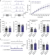
Figure 4. Deleting Ube3a in TH-positive neurons decreases GABA co-release and enhances motivational behaviour.
(a) Average nose pokes for inactive and active ports triggering 30 Hz optical intracranial self-stimulation in a 60 min behavioural session (n=5 and 7, one-way ANOVA *P<0.02). Experimental design was similar to that schematized in Fig. 1a, except that THCRE::Ube3aFLOX/p+ and THCRE::Ube3am+/p+ mice were examined to selectively delete Ube3a and optically stimulate TH+ VTA-to-NAc terminals. (b) Representative fast-scan cyclic voltammograms assessing dopamine release within the ventral striatum of THCRE::Ube3aFLOX/p+ and THCRE::Ube3am+/p+ mice. Dopamine release was evoked by 30 Hz (5-pulses) optical stimulation. Insets represent background-subtracted electrochemical signal characteristic of oxidized dopamine. (c) Averaged optically evoked dopamine release at a range of frequencies demonstrates that there are no statistical differences between genotypes (n=6 and 7, one-way ANOVA P>0.05). (d) Schematic representing protocol for whole-cell optical IPSC (oIPSC) recordings within the NAc of THCRE::Ube3aFLOX/p+ mice. IPSCs were pharmacologically isolated (see the Methods section). (e) Representative oIPSC traces measured in THCRE::Ube3aFLOX/p+ and THCRE::Ube3am+/p+ mice evoked by single pulses (20 ms) or 30 Hz trains of stimulation. The pharmacological isolation of GABAergic currents was validated by application of the antagonist SR95531. (f) Average peak amplitude of single oIPSCs demonstrate a significant reduction of GABAergic currents in Ube3aFLOX/p+ mice (n=13 and 15, **P<0.05). (g) Average decay kinetics of oIPSCs analysed in f (Student's t-test, P=0.61). Representative traces are shown with normalized amplitude to demonstrate similar current decay kinetics. (h) Average oIPSC charge evoked using a similar 30 Hz optical stimulation paradigm used behaviourally (see a) and tested in vitro (bottom traces of e). THCRE::Ube3aFLOX/p+ mice showed a significant decrease in GABA co-release (n=10 and 14, Student's t-test **P<0.05). All bars represent the mean±s.e.m.
Figure 5
Figure 5. Exogenous VGAT expression in THCRE-positive neurons normalizes motivation and GABA co-release in THCRE::Ube3aFLOX/p+ mice.
(a) Schematic representation of the experimental paradigm. DIO-VGAT and DIO-ChR2-eYFP were injected in experimental mice to selectively delete Ube3a in TH-expressing neurons and to express exogenous ChR2 and VGAT in TH+ VTA-to-NAc terminals. (b) Representative oIPSC evoked by single pulses (20 ms) or 30 Hz trains of stimulation. (c) Average oIPSC peak amplitudes evoked by single pulses (n=10 and 7, Student's t-test P=0.69), and (d) average total charge evoked by 30 Hz stimulation (n=7 and 7, Student's t-test P=0.59) demonstrate that exogenous VGAT expression in THCRE neurons normalizes GABA co-release between genotypes. (e) Response plots showing cumulative nose pokes that trigger a 30 Hz, 473 nm stimulus (active nose pokes) in representative mice. (f) Average nose pokes for inactive and active ports triggering 30 Hz optical intracranial self-stimulation in a 60 min behavioural session (n=6 and 6, one-way ANOVA P=0.12). Bars represent mean±s.e.m.

Similar articles

Cited by

References

    1. Schultz W. Predictive reward signal of dopamine neurons. J. Neurophysiol. 80, 1–27 (1998). - PubMed
    1. Schultz W. The phasic reward signal of primate dopamine neurons. Adv. Pharmacol. 42, 686–690 (1998). - PubMed
    1. Yun I. A., Wakabayashi K. T., Fields H. L. & Nicola S. M. The ventral tegmental area is required for the behavioral and nucleus accumbens neuronal firing responses to incentive cues. J. Neurosci. 24, 2923–2933 (2004). - PMC - PubMed
    1. Tritsch N. X. & Sabatini B. L. Dopaminergic modulation of synaptic transmission in cortex and striatum. Neuron 76, 33–50 (2012). - PMC - PubMed
    1. Tritsch N. X., Oh W. J., Gu C. & Sabatini B. L. Midbrain dopamine neurons sustain inhibitory transmission using plasma membrane uptake of GABA, not synthesis. Elife 3, e01936 (2014). - PMC - PubMed

Publication types

MeSH terms

LinkOut - more resources