Review
. 2016 Jan 28:10:189-206.
doi: 10.2147/OPTH.S80490. eCollection 2016.

Minimally invasive glaucoma surgery: current status and future prospects

Affiliations
Review

Minimally invasive glaucoma surgery: current status and future prospects

Grace M Richter et al. Clin Ophthalmol. .

Abstract

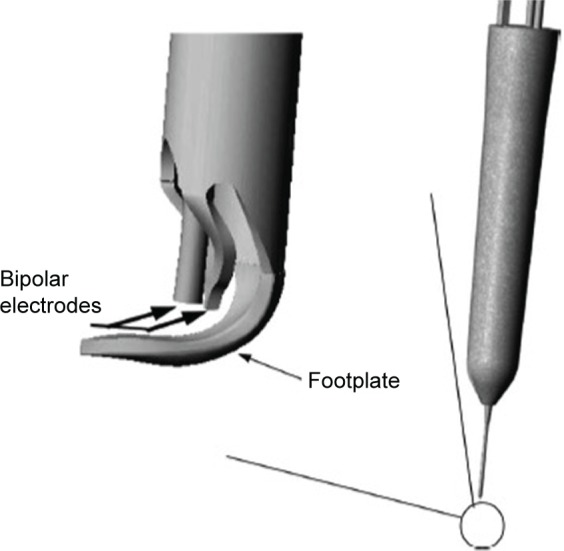
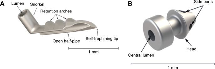
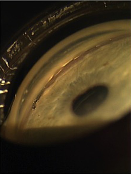
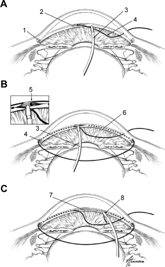
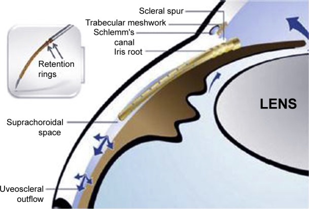
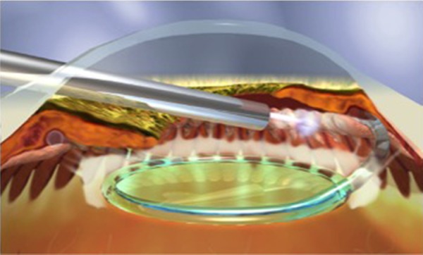
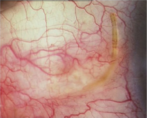
Minimally invasive glaucoma surgery aims to provide a medication-sparing, conjunctival-sparing, ab interno approach to intraocular pressure reduction for patients with mild-to-moderate glaucoma that is safer than traditional incisional glaucoma surgery. The current approaches include: increasing trabecular outflow (Trabectome, iStent, Hydrus stent, gonioscopy-assisted transluminal trabeculotomy, excimer laser trabeculotomy); suprachoroidal shunts (Cypass micro-stent); reducing aqueous production (endocyclophotocoagulation); and subconjunctival filtration (XEN gel stent). The data on each surgical procedure for each of these approaches are reviewed in this article, patient selection pearls learned to date are discussed, and expectations for the future are examined.

Keywords: MIGS; Schlemm’s canal; ab interno; microincisional glaucoma surgery; suprachoroidal shunt; trabecular stent.


Figures

Figure 1
Figure 1
Trabectome handpiece showing the footplate which glides within Schlemm’s canal and the bipolar electrodes which cauterize the inner wall of Schlemm’s canal. Note: Reproduced from Francis BA, See RF, Rao NA, Minckler DS, Baerveldt G. Ab interno trabeculectomy: development of a novel device (Trabectome) and surgery for open-angle glaucoma. J Glaucoma. 2006;15:68–73. Copyright © 2006. Promotional and commercial use of the material in print, digital or mobile device format is prohibited without the permission from the publisher Wolters Kluwer Health. Please contact healthpermissions@wolterskluwer.com for further information.
Figure 2
Figure 2
The first-generation trabecular iStent (A) with the self-trephining tip that is inserted into Schlemm’s canal via a sideways sliding technique, and then is maintained by the retention arches. The lumen is the portion facing the anterior chamber. The second-generation trabecular iStent inject (B), with four side ports within the Schlemm’s canal. The central lumen faces the anterior chamber. This model eliminates the need for the sideways sliding movement in the surgery technique. Note: Copyright © 2014. Dove Medical Press. Adapted from Hunter KS, Fjield T, Heitzmann H, Shandas R, Kahook MY. Characterization of micro-invasive trabecular bypass stents by ex vivo perfusion and computational flow modeling. Clin Ophthalmol. 2014;8:499–506.
Figure 3
Figure 3
The Hydrus trabecular stent sitting within a dilated Schlemm’s canal. Note: Reprinted from Grover DS, Godfrey DG, Smith O, Feuer WJ, Montes de Oca I, Fellman RL. Gonioscopy-assisted transluminal trabeculotomy, ab interno trabeculotomy: technique report and preliminary results. Ophthalmology. 2014; 121:855–861. Copyright © 2014, with permission from Elsevier.
Figure 4
Figure 4
Demonstration of gonioscopy-assisted transluminal trabeculotomy. Notes: An illuminated microcatheter is inserted through a nasal paracentesis wound (A). After a small goniotomy wound is created, the microcatheter is inserted into Schlemm’s canal using microsurgical forceps. After being guided around 360° (B), each end of the microcatheter is externalized from the temporal wound, creating a 360° trabeculotomy (C). 1=Schlemm’s canal; 2=initial goniotomy site; 3=microsurgical forceps; 4=microcatheter; 5=distal end of microcatheter after being passed 360° around Schlemm’s canal; 6=path of microcatheter within Schlemm’s canal; 7=trabecular shelf created after this procedure; 8=trabeculotomy created when the distal end of catheter is externalized. Reprinted from Pfeiffer N, Garcia-Feijoo J, Martinez-de-la-Casa JM, et al. A randomized trial of a Schlemm’s canal microstent with phacoemulsification for reducing intraocular pressure in open-angle glaucoma. Ophthalmology. 2015;122:1283–1293. Copyright © 2015, with permission from Elsevier.
Figure 5
Figure 5
The Cypass suprachoroidal shunt in position in the suprachoroidal space with retention rings near the ciliary body face. Notes: The blue arrows demonstrate the directional flow of aqueous. Reprinted from Hoeh H, Ahmed IK, Grisanti S, et al. Early postoperative safety and surgical outcomes after implantation of a suprachoroidal micro-stent for the treatment of open-angle glaucoma concomitant with cataract surgery. J Cataract Refract Surg. 2013;39:431–437. Copyright © 2013, with permission from Elsevier.
Figure 6
Figure 6
Endocyclophotocoagulation being performed on a pseudophakic patient. Note: Copyright © 2015. John Wiley and Sons. Reproduced from Siegel MJ, Boling WS, Faridi OS, et al. Combined endoscopic cyclophotocoagulation and phacoemulsification versus phacoemulsification alone in the treatment of mild to moderate glaucoma. Clin Experiment Ophthalmol. 2015;43:531–539.
Figure 7
Figure 7
External view of the XEN subconjunctival gel stent in place. Note: Reprinted from Lewis RA. Ab interno approach to the subconjunctival space using a collagen glaucoma stent. J Cataract Refract Surg. 2014;40:1301–1306. Copyright © 2014 with permission from Elsevier.

References

    1. Tham YC, Li X, Wong TY, Quigley HA, Aung T, Cheng CY. Global prevalence of glaucoma and projections of glaucoma burden through 2040: a systematic review and meta-analysis. Ophthalmology. 2014;121:2081–2090. - PubMed
    1. Heijl A, Leske MC, Bengtsson B, Hyman L, Bengtsson B, Hussein M, Early Manifest Glaucoma Trial Group Reduction of intraocular pressure and glaucoma progression: results from the Early Manifest Glaucoma Trial. Arch Ophthalmol. 2002;120:1268–1279. - PubMed
    1. Lemij HG, Hoevenaars JG, van der Windt C, Baudouin C. Patient satisfaction with glaucoma therapy: reality or myth? Clin Ophthalmol. 2015;9:785–793. - PMC - PubMed
    1. Leahy KE, White AJ. Selective laser trabeculoplasty: current perspectives. Clin Ophthalmol. 2015;9:833–841. - PMC - PubMed
    1. Woo DM, Healey PR, Graham SL, Goldberg I. Intraocular pressure-lowering medications and long-term outcomes of selective laser trabeculoplasty. Clin Experiment Ophthalmol. 2015;43:320–327. - PubMed