

Affective preclinical modeling of psychiatric disorders: taking imbalanced primal emotional feelings of animals seriously in our search for novel antidepressants
- PMID: 26869838
- PMCID: PMC4734875
- DOI: 10.31887/DCNS.2015.17.4/jpanksepp
Affective preclinical modeling of psychiatric disorders: taking imbalanced primal emotional feelings of animals seriously in our search for novel antidepressants
Abstract
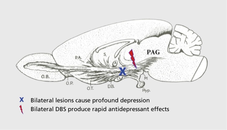
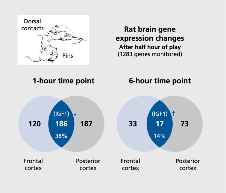
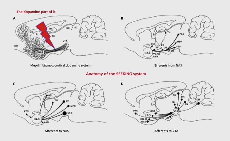
Preclinical animal models of psychiatric disorders are of critical importance for advances in development of new psychiatric medicine. Regrettably, behavior-only models have yielded no novel targeted treatments during the past half-century of vigorous deployment. This may reflect the general neglect of experiential aspects of animal emotions, since affective mental states of animals supposedly cannot be empirically monitored. This supposition is wrong-to the extent that the rewarding and punishing aspects of emotion circuit arousals reflect positive and negative affective states. During the past decade, the use of such affective neuroscience-based animal modeling has yielded three novel antidepressants (i) via the alleviation of psychic pain with low doses of buprenorphine; (ii) via the amplification of enthusiasm by direct stimulation of the medial forebrain bundle); and (iii) via the facilitation of the capacity for social joy with play facilitators such as rapastinel (GLYX13). All have progressed to successful human testing. For optimal progress, it may be useful for preclinical investigators to focus on the evolved affective foundations of psychiatrically relevant brain emotional disorders for optimal animal modeling.
Los modelos animales preclínicos de los trastornos psiquiátricos son de gran importancía para el avance en el desarrollo de la nueva medicina psiquiátrica. A pesar del importante desarrollo durante la segunda mitad del siglo pasado, los modelos puramente conductuales no han dado origen a nuevos blancos terapéuticos. Esto puede reflejar el rechazo general a los aspectos experienciales de las emocíones en los animales, ya que los estados mentales afectivos de ellos supuestamente no se pueden monitorear empíricamente. Esta suposición es incorrecta, dado que los aspectos de recompensa y castigo que activan los circuítos de las emociones reflejan estados afectivos positivos y negativos. Durante la última década el empleo de estos modelos animales basados en la neurociencia-afectiva ha producido tres nuevos antidepresivos: 1) aliviando el dolor psíquico con bajas dosis de buprenorfina, 2) amplificando el entusiasmo por estimulación directa del haz medial del cerebro anterior y 3) facilitando la capacidad de goce social con facilitadores del juego como el rapastinel (GLYX-13). Todos han avanzado con pruebas exitosas en humanos. Para un desarrollo óptimo, podría ser útil para los investigadores preclínicos enfocarse en las evolucion adas bases afectivas de los trastornos cerebrales emocionales importantes en psíquíatría para generar óptimos modelos animales.
Les modèles animaux précliniques de troubles psychiatriques sont d'une importance cruciale pour les avancées dans le développement de nouveaux médicaments psychiatriques. Malheureusement, durant ces 50 dernières années d'essor dynamique, aucun nouveau médicament ciblé n'est né de modèles fondés sur le seul comportement. Cela traduit peut-être le fait que les expériences négligent généralement les émotions animales, les états mentaux affectifs animaux n'étant pas supposés contrôlables empiriquement. Cette hypothèse est fausse dans la mesure où les dimensions de récompense et de punition de l'excitation des circuits de l'émotion reflètent des états affectifs positifs et négatifs. Ces 10 dernières années, trois nouveaux antidépresseurs sont issus de modèles animaux fondés sur cette neuroscience affective: 1) en soulageant la douleur psychique par de faibles doses de buprénorphine ; 2) en amplifiant l'enthousiasme par stimulation directe du faisceau médian du téléncéphale ; et 3) en facilitant la capacité de joie sociale par le biais de facilitateurs de jeu tels le rapastinel (GLYX-13). Ils ont tous franchi avec succès les étapes jusqu'aux essais chez l'homme. Pour un progrès maximal, les chercheurs en recherche préclinique devraient peut-être s'intéresser aux bases affectives évoluées des troubles émotionnels cérébraux psychiatriquement appropriés pour une modélisation animale optimale.
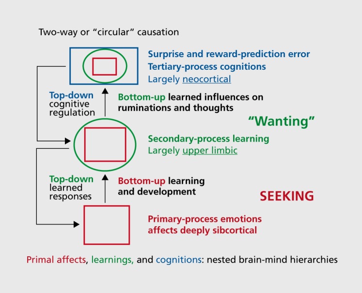
Keywords: antidepressant agent; buprenorphine; deep brain stimulation; emotional/affective behavior; medial forebrain bundle; primary process; secondary process; tertiary process.
Figures

Similar articles
-
The cross-mammalian neurophenomenology of primal emotional affects: From animal feelings to human therapeutics.J Comp Neurol. 2016 Jun 1;524(8):1624-35. doi: 10.1002/cne.23969. Epub 2016 Feb 15. J Comp Neurol. 2016. PMID: 26876723 Review.
-
Preclinical modeling of primal emotional affects (Seeking, Panic and Play): gateways to the development of new treatments for depression.Psychopathology. 2014;47(6):383-93. doi: 10.1159/000366208. Epub 2014 Oct 22. Psychopathology. 2014. PMID: 25341411 Review.
-
Cross-species affective neuroscience decoding of the primal affective experiences of humans and related animals.PLoS One. 2011;6(9):e21236. doi: 10.1371/journal.pone.0021236. Epub 2011 Sep 7. PLoS One. 2011. PMID: 21915252 Free PMC article. Review.
-
The Psycho-Neurology of Cross-Species Affective/Social Neuroscience: Understanding Animal Affective States as a Guide to Development of Novel Psychiatric Treatments.Curr Top Behav Neurosci. 2017;30:109-125. doi: 10.1007/7854_2016_458. Curr Top Behav Neurosci. 2017. PMID: 27696337
-
Toward a cross-species neuroscientific understanding of the affective mind: do animals have emotional feelings?Am J Primatol. 2011 Jun;73(6):545-61. doi: 10.1002/ajp.20929. Epub 2011 Feb 11. Am J Primatol. 2011. PMID: 21319205 Review.
Cited by
-
The anterior perforated substance (APS) revisited: Commented anatomical and imagenological views.Brain Behav. 2023 Dec;13(12):e3029. doi: 10.1002/brb3.3029. Epub 2023 Nov 27. Brain Behav. 2023. PMID: 38010896 Free PMC article.
-
Selected Principles of Pankseppian Affective Neuroscience.Front Neurosci. 2019 Jan 17;12:1025. doi: 10.3389/fnins.2018.01025. eCollection 2018. Front Neurosci. 2019. PMID: 30705615 Free PMC article. Review.
-
Affective Features Underlying Depression in Addiction: Understanding What It Feels Like.Front Psychol. 2019 Oct 17;10:2318. doi: 10.3389/fpsyg.2019.02318. eCollection 2019. Front Psychol. 2019. PMID: 31681110 Free PMC article.
-
Is panic disorder a disorder of physical fitness? A heuristic proposal.F1000Res. 2018 Mar 8;7:294. doi: 10.12688/f1000research.12788.1. eCollection 2018. F1000Res. 2018. PMID: 29623195 Free PMC article. Review.
-
Primary Emotional Systems and Personality: An Evolutionary Perspective.Front Psychol. 2017 Apr 11;8:464. doi: 10.3389/fpsyg.2017.00464. eCollection 2017. Front Psychol. 2017. PMID: 28443039 Free PMC article.
References
-
- Panksepp J. The vicissitudes of preclinical psychiatric research: justified abandonment by big pharma? Future Neurol. 2012;7(2):113–115.
-
- Markou A. Animal models of depression and antidepressant activity. Neurosci Biobehav Rev. 2005;29(4-5):501. - PubMed
-
- Watson JB. Behaviorism. New York, NY: W. W. Norton; 1929
Publication types
MeSH terms
Substances
LinkOut - more resources
Full Text Sources
Medical
Miscellaneous