. 2016 Feb 2:9:2.
doi: 10.3389/fnmol.2016.00002. eCollection 2016.

A Toolkit for Orthogonal and in vivo Optical Manipulation of Ionotropic Glutamate Receptors

Affiliations

A Toolkit for Orthogonal and in vivo Optical Manipulation of Ionotropic Glutamate Receptors

Joshua Levitz et al. Front Mol Neurosci. .

Abstract

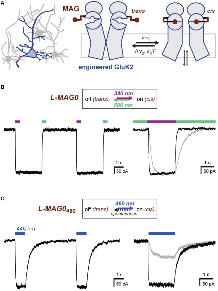
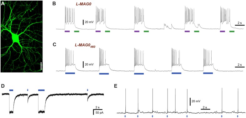
The ability to optically manipulate specific neuronal signaling proteins with genetic precision paves the way for the dissection of their roles in brain function, behavior, and disease. Chemical optogenetic control with photoswitchable tethered ligands (PTLs) enables rapid, reversible and reproducible activation or block of specific neurotransmitter-gated receptors and ion channels in specific cells. In this study, we further engineered and characterized the light-activated GluK2 kainate receptor, LiGluR, to develop a toolbox of LiGluR variants. Low-affinity LiGluRs allow for efficient optical control of GluK2 while removing activation by native glutamate, whereas variant RNA edited versions enable the synaptic role of receptors with high and low Ca(2+) permeability to be assessed and spectral variant photoswitches provide flexibility in illumination. Importantly, we establish that LiGluR works efficiently in the cortex of awake, adult mice using standard optogenetic techniques, thus opening the door to probing the role of specific synaptic receptors and cellular signals in the neural circuit operations of the mammalian brain in normal conditions and in disease. The principals developed in this study are widely relevant to the engineering and in vivo use of optically controllable proteins, including other neurotransmitter receptors.

Keywords: chemical optogenetics; glutamate receptor; in vivo; molecular engineering; photo-pharmacology.


Figures

Figure 1
Figure 1
Operating principles of light-gated glutamate receptors (LiGluRs). (A) LiGluRs are optogenetic tools that can be expressed in genetically targeted cells (blue). They are based on an engineered iGluR subunit, which is expressed in the desired cell type and conjugated with a synthetic photoswitchable ligand, called MAG. Photoisomerization of the covalently bound MAG ligand from trans to cis presents the glutamate moiety to the glutamate binding site, which leads to ligand binding and ion channel opening. (B) Photoswitching of LiGluR labeled with a regular, bistable MAG ligand (Volgraf et al., ; Gorostiza et al., 2007). Illumination with 380 nm light (violet bar) leads to an inward current as shown in a voltage-clamp recording of a LiGluR-expressing HEK cell labeled with L-MAG0. The receptor activation is sustained in the dark and turned off by illumination with 500 nm light (green bar). (Left) Switching is fully reversible as demonstrated with two consecutive switching cycles. (Right) Lowering the light intensity leads to slower photo-activation and deactivation kinetics, but the same current amplitude (black trace ~7-8 mW/mm2; gray trace ~0.7-0.8 mW/mm2; see also Figure S2). (C) Photoswitching of LiGluR labeled with L-MAG0460,a blue light activated photoswitch with a fast spontaneous cis-to-trans relaxation (Kienzler et al., 2013). (Left) HEK cell voltage-clamp recording showing two switching cycles with 445 nm light (blue bar). Once the blue light is turned off, LiGluR turns off spontaneously. (Right) Lowering the light intensity results in slower activation kinetics and a decreased response (black trace ~1.5 mW/mm2; gray trace ~0.1 mW/mm2). HEK cell recordings were performed in the presence of ConA.
Figure 2
Figure 2
Light-induced depolarization and optical control of neuronal firing with LiGluR. (A) LiGluR-GFP expression in a cultured hippocampal neuron visualized with confocal imaging. Scale bar = 20 μm. (B) LiGluR labeling with conventional L-MAG0 allows for optical depolarization in a bistable fashion: 375 nm light (violet bar) depolarizes the cells, which results in an increase in action potential firing, illumination with 480 nm light (green bar) readily reverses the effect (current-clamp recording at Vrest = −60 mV). (C–E) LiGluR labeled with L-MAG0460 allows optical control of neuronal firing with light of a single, blue wavelength. (C) The effect of 445 nm illumination (blue bar) reverses spontaneously in the dark (current-clamp recording at Vrest = −57 mV). (D) Voltage-clamp recording at −60 mV showing inward currents resulting from long (seconds) and short (5 ms) 445 nm light pulses. (E) Short 445 nm light pulses (arrows; 3 ms) are sufficient to elicit single action potentials (current-clamp recording at Ubase = −47 mV).
Figure 3
Figure 3
Engineering and characterization of LiGluR variants with low glutamate affinity. (A) Glutamate dose-response properties of LiGluR, LA-LiGluR [LiGluR(K487A)] and ULA-LiGluR [LiGluR(E738D)] obtained from HEK cell voltage-clamp recordings in the presence of Con A. Apparent affinities were determined by fitting with a Hill-type equation, which yielded EC50 = (0.70 ± 0.16) mM (Θ = 1.00± 0.07, n = 3 cells) for LA-LiGluR, and EC50 = (10.0 ± 2.6) mM (Θ = 0.95 ± 0.10, n = 4 cells) for ULA-LiGluR. Data points report the mean ± s.d. LiGluR was reported to have a glutamate EC50 = (52 ± 1) μM (from Gorostiza et al., 2007). (B) Photoactivation of LA-LiGluR after labeling with regular L-MAG0 (top) and L-MAG0460 (bottom). Voltage-clamp recordings in the presence of ConA with DG4 illumination (1–2 mW/mm2), (C) Confocal image of hippocampal neurons expressing LA-LiGluR-EGFP. Scale bars = 10 μm. (D) Optical control of hippocampal neurons using LA-LiGluR-EGFP labeled with regular L-MAG0 (current-clamp recording at Vrest = −76 mV), or, (E) labeled with L-MAG0460 (current-clamp recording at Vbase = −40 mV). (F) Summary of light-induced depolarization achieved with different combinations of LiGluR and MAG from a common potential of −60 mV (mean ± s.d. with number of cells). An ANOVA test did not detect significant differences between the groups (n.s.).
Figure 4
Figure 4
In vivo optical control of LiGluR in the visual cortex of awake mice. (A) Top, map for AAV vector used to produce AAV-hsyn-LiGluR. Bottom, expression of GluK2/LiGluR in V1 neurons as visualized by immunohistochemistry. Scale bars = 200 μm, left and 10 μm, right. (B) Schematic showing experimental setup of in vivo electrophysiology experiments. A TTL-controlled dual laser system was connected to an optrode which was implanted into V1 of an awake, head-fixed mouse and was attached to a TDT-Rz5 amplifier. (C,D) Representative recordings from either a LiGluR (C) or GFP (D) injected mouse following L-MAG0 injection. Top, unfiltered recordings of electrical activity in response to 375 nm (violet) or 532 nm (green) illumination. Bottom, high-pass filtered electrical recordings. (E,F), Representative raster plots for individual units showing repeatability of LiGluR photoactivation (E) and no response in neurons not infected with LiGluR (F). (G) Local field potential (LFP) responses to LiGluR activation were observed for L-MAG0. Data is presented as normalized power (z-axis, color coded) as a function of time (x-axis) and frequency (y-axis). Power was normalized to the 1 s period prior to laser stimulation for each frequency. (H) No LFP response was observed in GFP-injected mice.
Figure 5
Figure 5
Further characterization of in vivo optical control of LiGluR. (A) Summary of light response from a representative cell showing the speed of response. (B) Kinetics for the activation (purple) and inactivation (green) of LiGluR with L-MAG0. Values represent the delay between the onset of laser stimulation and the earliest change in firing rate, averaged across all single units recorded. Data is presented as mean ± s.e.m. (C) Representative filtered recording from V1 of a LiGluR-expressing and L-MAG0-labeled mouse showing high frequency light responses. (D) Summary of firing rate changes in a representative cell in response to high frequency photoswitching. (E) Summary histogram for all units recorded from either LiGluR (black) or GFP (gray) -infected mice. (F) Baseline firing rates (y-axis, mean ± s.e.m.) for periods between laser stimulations, in mice expressing LiGluR or GFP. No difference was observed between the two groups.
Figure 6
Figure 6
Single wavelength optical control of LiGluR in vivo with L-MAG0460. (A,B) LiGluR activation with L-MAG0460 results in increased firing in response to 473 nm light that is rapid, repeatable and spontaneously-reversed in the dark. (C) LFP changes are observed in response to LiGluR activation with L-MAG0460. (D) Kinetics for the activation (blue) and inactivation (gray) of LiGluR using L-MAG0460. (E) Representative recording showing power-dependence of LiGluR photoactivation with L-MAG0460. (F) Summary of power-dependence of photoactivation for a representative cell. (G,H) 24 h post MAG injection light responses to 473 nm light were maintained.

Similar articles

Cited by

References

    1. Anikeeva P., Andalman A. S., Witten I., Warden M., Goshen I., Grosenick L., et al. . (2012). Optetrode: a multichannel readout for optogenetic control in freely moving mice. Nat. Neurosci. 15, 163–170. 10.1038/nn.2992 - DOI - PMC - PubMed
    1. Banghart M., Borges K., Isacoff E., Trauner D., Kramer R. H. (2004). Light-activated ion channels for remote control of neuronal firing. Nat. Neurosci. 7, 1381–1386. 10.1038/nn1356 - DOI - PMC - PubMed
    1. Banghart M. R., Volgraf M., Trauner D. (2006). Engineering light-gated ion channels. Biochemistry 45, 15129–15141. 10.1021/bi0618058 - DOI - PubMed
    1. Broichhagen J., Damijoinatas A., Levitz J., Sokol K., Leippe P., Konrad D., et al. (2015). Orthogonal optical control of a class C G protein-coupled receptor using a SNAP-tethered photoswitch. ACS Central Sci. 1, 383–393. 10.1021/acscentsci.5b00260 - DOI - PMC - PubMed
    1. Browne L. E., Nunes J. P., Sim J. A., Chudasama V., Bragg L., Caddick S., et al. . (2014). Optical control of trimeric P2X receptors and acid-sensing ion channels. Proc. Natl. Acad. Sci. U.S.A. 111, 521–526. 10.1073/pnas.1318582111 - DOI - PMC - PubMed