. 2016 Feb 3:9:7.
doi: 10.3389/fnmol.2016.00007. eCollection 2016.

Expression of Tgfβ1 and Inflammatory Markers in the 6-hydroxydopamine Mouse Model of Parkinson's Disease

Affiliations

Expression of Tgfβ1 and Inflammatory Markers in the 6-hydroxydopamine Mouse Model of Parkinson's Disease

Stefan Jean-Pierre Haas et al. Front Mol Neurosci. .

Abstract

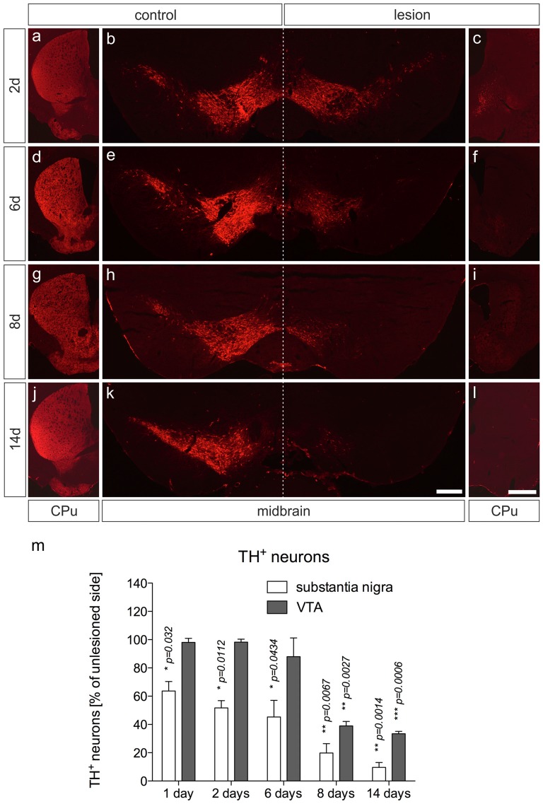
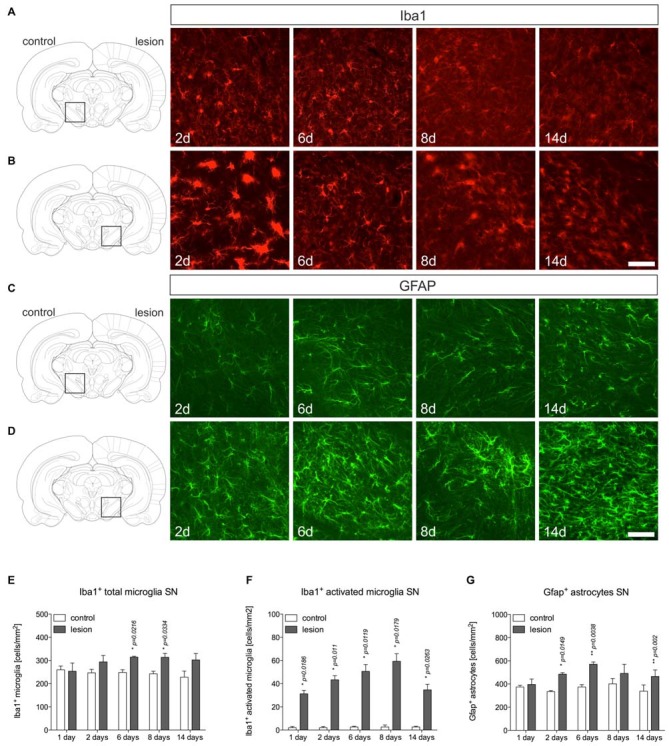
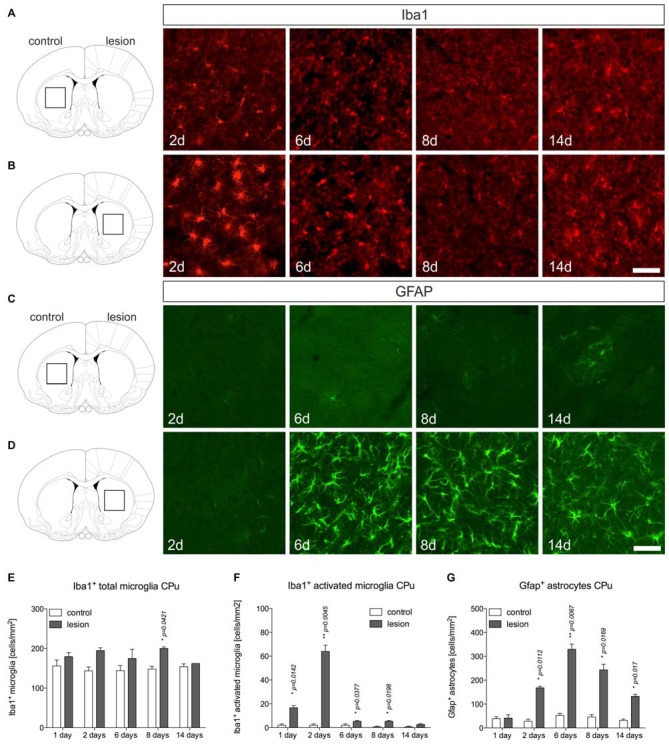
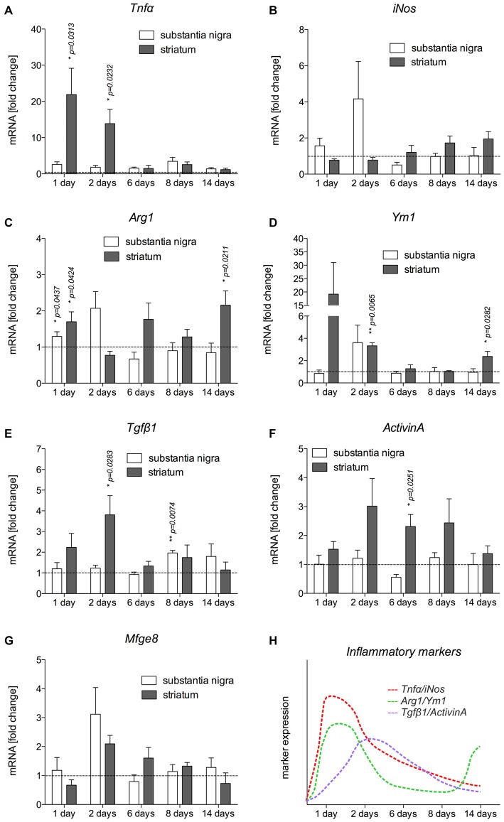
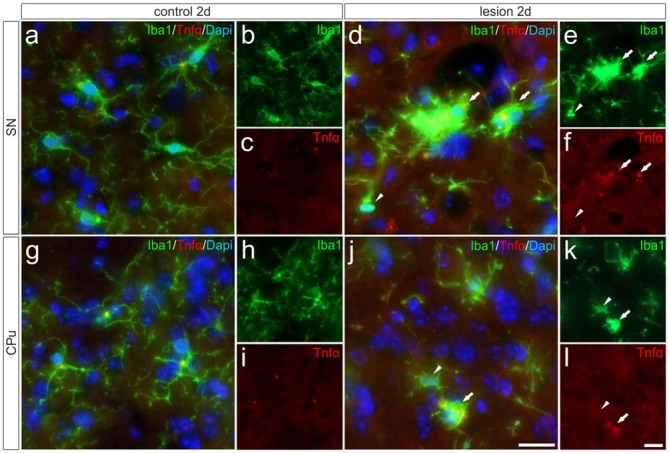
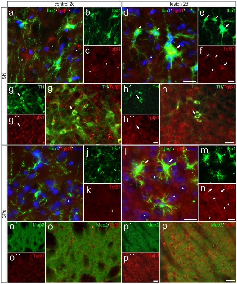
Parkinson's disease (PD) is a neurodegenerative disorder that is characterized by loss of midbrain dopaminergic (mDA) neurons in the substantia nigra (SN). Microglia-mediated neuroinflammation has been described as a common hallmark of PD and is believed to further trigger the progression of neurodegenerative events. Injections of 6-hydroxydopamine (6-OHDA) are widely used to induce degeneration of mDA neurons in rodents as an attempt to mimic PD and to study neurodegeneration, neuroinflammation as well as potential therapeutic approaches. In the present study, we addressed microglia and astroglia reactivity in the SN and the caudatoputamen (CPu) after 6-OHDA injections into the medial forebrain bundle (MFB), and further analyzed the temporal and spatial expression patterns of pro-inflammatory and anti-inflammatory markers in this mouse model of PD. We provide evidence that activated microglia as well as neurons in the lesioned SN and CPu express Transforming growth factor β1 (Tgfβ1), which overlaps with the downregulation of pro-inflammatory markers Tnfα, and iNos, and upregulation of anti-inflammatory markers Ym1 and Arg1. Taken together, the data presented in this study suggest an important role for Tgfβ1 as a lesion-associated factor that might be involved in regulating microglia activation states in the 6-OHDA mouse model of PD in order to prevent degeneration of uninjured neurons by microglia-mediated release of neurotoxic factors such as Tnfα and nitric oxide (NO).

Keywords: 6-OHDA; Tgfβ1; Tnfα; astrocytes; microglia.


Figures

Figure 1
Figure 1
6-OHDA injections into the MFB induce robust loss of mDA neurons and denervation in the CPu on the lesioned side. Coronal brain sections after immunohistochemistry for TH from CPu and ventral midbrains containing SN and VTA are shown 2 days (A–C), 6 days (D–F), 8 days (G–I) and 14 days (J–L) after 6-OHDA injections. Whereas loss of TH+ fibers in the CPu occurs from 2 days onwards, substantial decrease in numbers of TH+ mDA neurons was evident after 6 days. Representative images from three independent experiments are shown. Scale bars indicate 300 μm (B,E,H,K) and 1 mm for all CPu images. Quantification of SN and VTA TH+ mDA neurons revealed significant decreases in SN neurons at all time-points analyzed. However, decrease of VTA neurons was delayed and reached significant differences at 8 days and 14 days (M). Data are given as percentages from unlesioned sides ± SEM from three animals.
Figure 2
Figure 2
Microglia and astroglia reactivity in the SN after 6-OHDA injections. On the control side microglia showing ramified morphologies could be detected by Iba1-immunohistochemistry at all time-points analyzed (A). Two days after 6-OHDA injections, a strong microglia reactivity was observed on the lesioned side. After 6 days, microglia start to adopt a ramified morphology indicating that microglia reactivity is rapidly inhibited (B). Immunohistochemistry for Gfap demonstrated the presence of Gfap+ astrocytes in the SN on the control side at all time-points analyzed (C). However, strong increases in Gfap-immunoreactivity and numbers of Gfap+ astrocytes were observed starting from 6 days after injection of 6-OHDA and persisting until 14 days (D). Scale bars indicate 75 μm. Quantifications of total microglia (E), activated microglia (F) and Gfap+ astrocytes (G) in the SN on control and lesioned sides. Data are given as numbers/mm2 (mean ± SEM) from three animals per time-point. p-values derived from student’s t-test are *p < 0.05 and **p < 0.01.
Figure 3
Figure 3
Microglia and astroglia reactivity in the CPu after 6-OHDA injections. Microglia showing ramified morphologies could be detected after Iba1-immunohistochemistry in the unlesioned CPu at all time-points analyzed (A). Two days after 6-OHDA injections a strong microglia reactivity was observed in the lesioned CPu, which was not longer visible at 6 days, 8 days and 14 days after injection of 6-OHDA (B). Immunohistochemistry for Gfap showed that virtually no Gfap+ astrocytes were present in the CPu on the control side at all time-points analyzed (C). Comparable to the observations in the lesioned SN, strong increases in Gfap-immunoreactivity and numbers of Gfap+ astrocytes in the CPu were observed starting from 6 days after injection of 6-OHDA and again, persisting until 14 days (D). Scale bars indicate 75 μm. Quantifications of total microglia (E), activated microglia (F) and Gfap+ astrocytes (G) in the CPu on control and lesioned sides. Data are given as numbers/mm2 (mean ± SEM) from three animals per time-point. p-values derived from student’s t-test are *p < 0.05 and **p < 0.01.
Figure 4
Figure 4
Expression of inflammatory markers in SN and CPu after injection of 6-OHDA. The expression levels of the M1 activation markers Tnfα (A) and iNos (B), the M2 activation markers Ym1 (C) and Arg1 (D) as well as TGFβ1 (E) and ActivinA (F) as anti-inflammatory factors as well as Mfge8 expression (G) as a TGFβ1-regulated gene have been analyzed and are displayed. Data are given as fold changes (mean ± SEM) calculated from expression levels on unlesioned control sides from at least three different animals. p-values derived from student’s t-test are *p < 0.05 and **p < 0.01. A summary of the temporal expression patterns of different markers is given (H).
Figure 5
Figure 5
Microglial expression of Tnfα in the SN und the CPu after 6-OHDA injections. Two days after 6-OHDA-induced lesion no Tnfα expression was observed in microglia in SN (A–C) and CPu (G–I). However, distinct Tnfα immunoreactivity could be detected on the lesioned side. Activated microglia (white arrows) showed Tnfα expression in the SN (D–F) as well as in the CPu (J–L). No Tnfα signals were present in ramified non-activated microglia (white arrowheads) from the SN (D–F) and the CPu (J–L) on the lesioned side. Representative images from three different animals are shown. Scale bars indicate 20 μm.
Figure 6
Figure 6
Microglial expression of Tgfβ1 in the SN und the CPu after 6-OHDA injections. Two days after 6-OHDA-induced lesion weak microglial and distinct neuronal Tgfβ1 expression (white asterisks) was observed in SN (A–C,G–G″) and CPu (I–K,O,O′). However, increased Tgfβ1 immunoreactivity could be detected on the lesioned side after 2 days (H–H″). Activated microglia (white arrows) showed Tgfβ1 expression in the SN (D–F), whereas ramified microglia (white arrowheads) showed no Tgfβ1 expression (D–F). A strong increase in Tgfβ1 immunoreactivity was observed in the lesioned CPu 2 days after 6-OHDA injections (L–N). Next to activated microglia (white arrows) and neurons (white asterisks) the neuropil displayed prominent Tgfβ1 signals in the lesioned CPu (L,N). Map2-positive striatal neurons displayed a weak Tgfβ1 immunoreactivity on control sides (O–O″). After lesion, strong Tgfβ1 signals could be observed in Map2-positive neurons in the CPu (P–P″). Representative images from three different animals are shown. Scale bars indicate 20 μm (A–F,I–N) and 50 μm (G,H,O,P).

References

    1. Barbeau A. (1969). L-Dopa therapy in Parkinson’s disease: a critical review of nine years’ experience. Can. Med. Assoc. J. 101, 59–68. - PMC - PubMed
    1. Block M. L., Zecca L., Hong J.-S. (2007). Microglia-mediated neurotoxicity: uncovering the molecular mechanisms. Nat. Rev. Neurosci. 8, 57–69. 10.1038/nrn2038 - DOI - PubMed
    1. Bortolanza M., Cavalcanti-Kiwiatkowski R., Padovan-Neto F. E., da Silva C. A., Mitkovski M., Raisman-Vozari R., et al. (2014). Glial activation is associated with l-DOPA induced dyskinesia and blocked by a nitric oxide synthase inhibitor in a rat model of Parkinson’s disease. Neurobiol. Dis. 73, 377–387. 10.1016/j.nbd.2014.10.017 - DOI - PubMed
    1. Butovsky O., Jedrychowski M. P., Moore C. S., Cialic R., Lanser A. J., Gabriely G., et al. (2014). Identification of a unique Tgf-β-dependent molecular and functional signature in microglia. Nat. Neurosci. 17, 131–143. 10.1038/nn.3599 - DOI - PMC - PubMed
    1. Chen L. W., Yung K. L., Chan Y. S. (2005). Reactive astrocytes as potential manipulation targets in novel cell replacement therapy of Parkinson’s disease. Curr. Drug Targets 6, 821–833. 10.2174/138945005774574506 - DOI - PubMed