. 2016 Feb 3:10:3.
doi: 10.3389/fncir.2016.00003. eCollection 2016.

Whole-Brain Mapping of Neuronal Activity in the Learned Helplessness Model of Depression

Affiliations

Whole-Brain Mapping of Neuronal Activity in the Learned Helplessness Model of Depression

Yongsoo Kim et al. Front Neural Circuits. .

Abstract

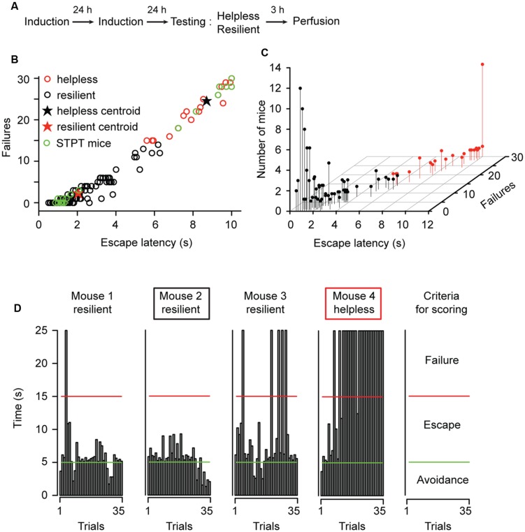
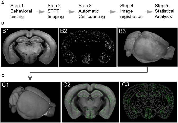
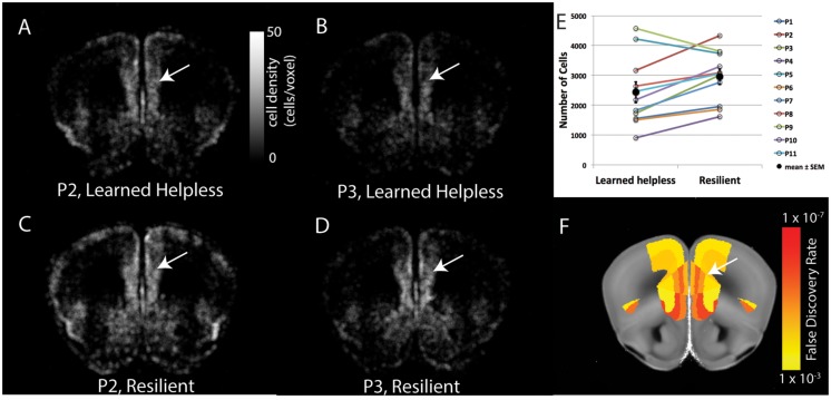
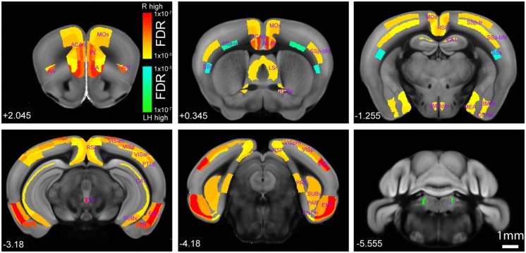
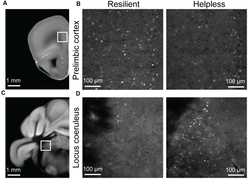
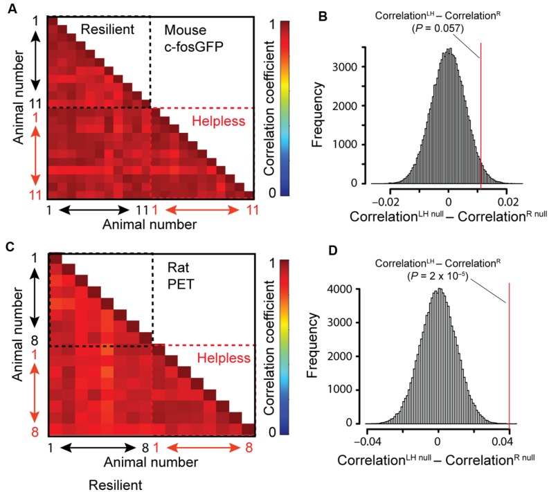
Some individuals are resilient, whereas others succumb to despair in repeated stressful situations. The neurobiological mechanisms underlying such divergent behavioral responses remain unclear. Here, we employed an automated method for mapping neuronal activity in search of signatures of stress responses in the entire mouse brain. We used serial two-photon tomography to detect expression of c-FosGFP - a marker of neuronal activation - in c-fosGFP transgenic mice subjected to the learned helplessness (LH) procedure, a widely used model of stress-induced depression-like phenotype in laboratory animals. We found that mice showing "helpless" behavior had an overall brain-wide reduction in the level of neuronal activation compared with mice showing "resilient" behavior, with the exception of a few brain areas, including the locus coeruleus, that were more activated in the helpless mice. In addition, the helpless mice showed a strong trend of having higher similarity in whole-brain activity profile among individuals, suggesting that helplessness is represented by a more stereotypic brain-wide activation pattern. This latter effect was confirmed in rats subjected to the LH procedure, using 2-deoxy-2[18F]fluoro-D-glucose positron emission tomography to assess neural activity. Our findings reveal distinct brain activity markings that correlate with adaptive and maladaptive behavioral responses to stress, and provide a framework for further studies investigating the contribution of specific brain regions to maladaptive stress responses.

Keywords: C-fos expression; Positron-emission tomography; depression; learned helplessness; serial two-photon tomography.


Figures

FIGURE 1
FIGURE 1
Characterization of behavioral responses in the learned helplessness procedure. (A) A schematic of the experimental procedure. (B) A graphic representation of the behavioral measurements (escape latency and failures) for all mice (n = 144) used in this study. A k-means cluster analysis defined two distinct clusters, the “helpless” and “resilient.” Mice chosen for further STPT imaging analysis is highlighted with green dots (STPT mice). (C) A 3D scatterplot of the same data shown in (B). The position of each dot along the z-axis represents the number of mice showing a specific set of behavioral parameters. (D) Behavioral measurements from four representative mice tested in parallel. Each graph shows the time (y-axis) an animal took to finish each of the 35 trials (x-axis) in a testing session (see Materials and Methods). Trials that were ended within 5 s (bars under the green line) represent avoidance; trials that were ended within 15 s (bars under the red line) represent escape. The remaining trials represent failure. The rightmost panel summarizes the criteria for scoring. Mouse 2 and mouse 4 (marked by a black box and red box, respectively) were mice with extreme behavioral measurements, and they were chosen for STPT imaging (total 11 pairs, with one helpless and one resilient in each pair).
FIGURE 2
FIGURE 2
Imaging whole-brain c-FosGFP in the c-FosGFP mice. (A) A schematic of the cFos-GFP whole-brain data processing pipeline. (B; B1) A 2D-image of one sample brain taken with STPT. (B2) Machine learning algorithm-based c-FosGFP detection from the image shown in B1. (B3) 3D-reconstruction of the sample brain from serial 2D-images, including the one shown in B1. (C; C1) The sample brain is registered to a reference STPT brain before statistical analysis. (C2) The c-FosGFP signal transformed onto the reference brain. (C3) The reference brain is equipped with Allen Reference Brain anatomical labeling (http://atlas.brain-map.org).
FIGURE 3
FIGURE 3
Pair-wise comparison of brain cFos-GFP expression between the helpless and the resilient mice. (A–D) The density of cFos-GFP+ cells in evenly spaced and overlapping sphere voxel (100 μm diameter) in a coronal brain section containing the prelimbic cortex, for two pairs of mice: P2 (A, helpless, C, resilient) and P3 (B, helpless, D, resilient). Heat map represents cFos-GFP cell counts per voxel. White arrows denote the prelimbic cortex. (E) Quantification of the number of cFos-GFP+ cells in the prelimbic cortex for the 11 pairs of mice. Filled circles and associated bars represent mean ± SEM. (F) The statistical results of pair-wise comparison for this brain section after multiple comparison correction by false discovery rate (FDR). Heat map represents FDR adjusted P-values. White arrow denotes the prelimbic cortex.
FIGURE 4
FIGURE 4
Brain-wide comparison between the helpless and resilient mice. Hot or cold colors represent anatomical regions of interest (ROI) with significantly higher activity in the resilient group (“R high”) or the learned helpless group (“LH high”), respectively, when comparisons are made between the two groups. Heat maps represent FDR q-values corrected for multiple comparisons after individual P-value in each brain region was computed. Numbers at the bottom denote coordinates relative to Bregma. A full dataset and the raw data containing cell counting in each anatomical area can be found in Supplementary Tables S1 and S2. Please also see Table 1. The Interactive Allen Reference atlas with anatomical annotations can be found in http://atlas.brain-map.org.
FIGURE 5
FIGURE 5
Raw STPT data from selected regions of the c-fosGFP mice. (A) A low magnification image of a brain section containing the prelimbic cortex (PL; boxed area). (B) High magnification images of the PL in a resilient mouse (left) and a helpless mouse (right). (C) A low magnification image of a brain section containing the locus coeruleus (LC; boxed area). (D) High magnification images of the LC in a resilient mouse (left) and a helpless mouse (right).
FIGURE 6
FIGURE 6
Correlation between individual animals in brain-wide activity. (A,B) Correlation analysis of c-FosGFP signal from the c-fosGFP mice. (A) Correlation coefficient for each pair of animals is displayed in a pairwise correlation matrix. (B) Result of the bootstrap test (see Materials and Methods for details). LH, learned helpless; R, resilient. Red line denotes the location in the null distribution (CorrelationLHnull – CorrelationRnull) where the difference between the actual LH and R groups (CorrelationLH – CorrelationR) fell (P-value resulted from a two-sided test). (C,D) Same as (A,B), except that correlation analysis was performed on PET data from rats.

References

    1. Abramson L. Y., Seligman M. E., Teasdale J. D. (1978). Learned helplessness in humans: critique and reformulation. J. Abnorm. Psychol. 87 49–74. 10.1037/0021-843X.87.1.49 - DOI - PubMed
    1. Bangasser D. A., Curtis A., Reyes B. A. S., Bethea T. T., Parastatidis I., Ischiropoulos H., et al. (2010). Sex differences in corticotropin-releasing factor receptor signaling and trafficking: potential role in female vulnerability to stress-related psychopathology. Mol. Psychiatry 15 877 896–904. 10.1038/mp.2010.66 - DOI - PMC - PubMed
    1. Barth A. L. (2004). Alteration of neuronal firing properties after in vivo experience in a FosGFP transgenic mouse. J. Neurosci. 24 6466–6475. 10.1523/JNEUROSCI.4737-03.2004 - DOI - PMC - PubMed
    1. Bradley A. J., Lenox-Smith A. J. (2013). Does adding noradrenaline reuptake inhibition to selective serotonin reuptake inhibition improve efficacy in patients with depression? A systematic review of meta-analyses and large randomised pragmatic trials. J. Psychopharmacol. 27 740–758. 10.1177/0269881113494937 - DOI - PubMed
    1. Chourbaji S., Zacher C., Sanchis-Segura C., Dormann C., Vollmayr B., Gass P. (2005). Learned helplessness: validity and reliability of depressive-like states in mice. Brain Res. Brain Res. Protoc. 16 70–78. 10.1016/j.brainresprot.2005.09.002 - DOI - PubMed

Publication types

MeSH terms

Substances