Review
. 2016 Feb 2:7:10.
doi: 10.3389/fphar.2016.00010. eCollection 2016.

Syndecans as Cell Surface Receptors in Cancer Biology. A Focus on their Interaction with PDZ Domain Proteins

Affiliations
Review

Syndecans as Cell Surface Receptors in Cancer Biology. A Focus on their Interaction with PDZ Domain Proteins

Bill Cheng et al. Front Pharmacol. .

Abstract

Syndecans are transmembrane receptors with ectodomains that are modified by glycosaminoglycan chains. The ectodomains can interact with a wide variety of molecules, including growth factors, cytokines, proteinases, adhesion receptors, and extracellular matrix (ECM) components. The four syndecans in mammals are expressed in a development-, cell-type-, and tissue-specific manner and can function either as co-receptors with other cell surface receptors or as independent adhesion receptors that mediate cell signaling. They help regulate cell proliferation and migration, angiogenesis, cell/cell and cell/ECM adhesion, and they may participate in several key tumorigenesis processes. In some cancers, syndecan expression regulates tumor cell proliferation, adhesion, motility, and other functions, and may be a prognostic marker for tumor progression and patient survival. The short cytoplasmic tail is likely to be involved in these events through recruitment of signaling partners. In particular, the conserved carboxyl-terminal EFYA tetrapeptide sequence that is present in all syndecans binds to some PDZ domain-containing proteins that may function as scaffold proteins that recruit signaling and cytoskeletal proteins to the plasma membrane. There is growing interest in understanding these interactions at both the structural and biological levels, and recent findings show their high degree of complexity. Parameters that influence the recruitment of PDZ domain proteins by syndecans, such as binding specificity and affinity, are the focus of active investigations and are important for understanding regulatory mechanisms. Recent studies show that binding may be affected by post-translational events that influence regulatory mechanisms, such as phosphorylation within the syndecan cytoplasmic tail.

Keywords: PDZ domain; cancer; cytoskeleton; extracellular matrix; phosphorylation; syndecan.


Figures
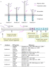
FIGURE 1
FIGURE 1
(A) The syndecan family of heparan sulfate proteoglycans. Syndecans consist of a transmembrane core protein with a short cytoplasmic tail and an ectodomain. All syndecan ectodomains are modified with either heparan sulfate only or heparan sulfate and chondroitin sulfate sugar chains. (B) A schematic showing the syndecan domains and examples of functions and interacting partners. (C) List of known PDZ-containing proteins that interact with syndecans.
FIGURE 2
FIGURE 2
(A) Structural definition of syndecan binding to the PDZ domain. Ribbon diagram of the syntenin PDZ2 bound to the syndecan-4 peptide TNEFYA (pdb code 1 OBY, Kang et al., 2003), (B) Surface charge (blue positive and red negative) representation of the PDZ2 domain with syndecan-4 (magenta) peptide displayed as ball-and-stick. (C) Similar representation of PDZ2 with a model of syndecan-1 tyrosine phosphorylated peptide (yellow) showed as ball-and-stick. (D) Phosphorylation of Tyr and Ser residues within syndecan cytoplasmic tails and their effects on PDZ protein binding: (-) no interaction; (+) interaction; (++) enhanced interaction. The conserved (C1 and C2) and variable (V) domain organization is from Couchman et al. (2015).

References

    1. Alexopoulou A. N., Multhaupt H. A., Couchman J. R. (2007). Syndecans in wound healing, inflammation and vascular biology. Int. J. Biochem. Cell Biol. 39 505–528. 10.1016/j.biocel.2006.10.014 - DOI - PubMed
    1. Altemeier W. A., Schlesinger S. Y., Buell C. A., Parks W. C., Chen P. (2012). Syndecan-1 controls cell migration by activating Rap1 to regulate focal adhesion disassembly. J. Cell Sci. 125 5188–5195. 10.1242/jcs.109884 - DOI - PMC - PubMed
    1. Anttonen A., Heikkilä P., Kajanti M., Jalkanen M., Joensuu H. (2001). High syndecan-1 expression is associated with favourable outcome in squamous cell lung carcinoma treated with radical surgery. Lung Cancer 32 297–305. 10.1016/S0169-5002(00)00230-0 - DOI - PubMed
    1. Asundi V. K., Carey D. J. (1995). Self-association of N-syndecan (syndecan-3) core protein is mediated by a novel structural motif in the transmembrane domain and ectodomain flanking region. J. Biol. Chem. 270 26404–26410. 10.1074/jbc.270.44.26404 - DOI - PubMed
    1. Asundi V. K., Carey D. J. (1997). Phosphorylation of recombinant N-Syndecan (Syndecan 3) core protein. Bioch. Biophys. Res. Comm. 240 502–506. 10.1006/bbrc.1997.7684 - DOI - PubMed

LinkOut - more resources