Review
. 2016 Feb 1:7:42.
doi: 10.3389/fmicb.2016.00042. eCollection 2016.

Bacterial Toxins as Pathogen Weapons Against Phagocytes

Affiliations
Review

Bacterial Toxins as Pathogen Weapons Against Phagocytes

Ana do Vale et al. Front Microbiol. .

Abstract

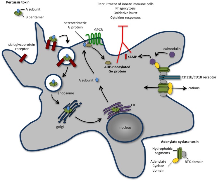
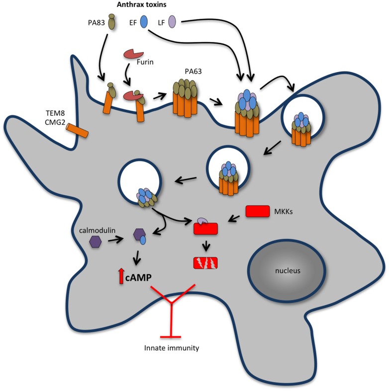
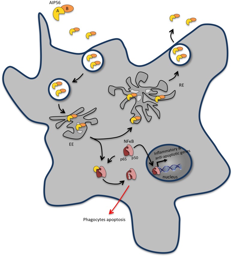
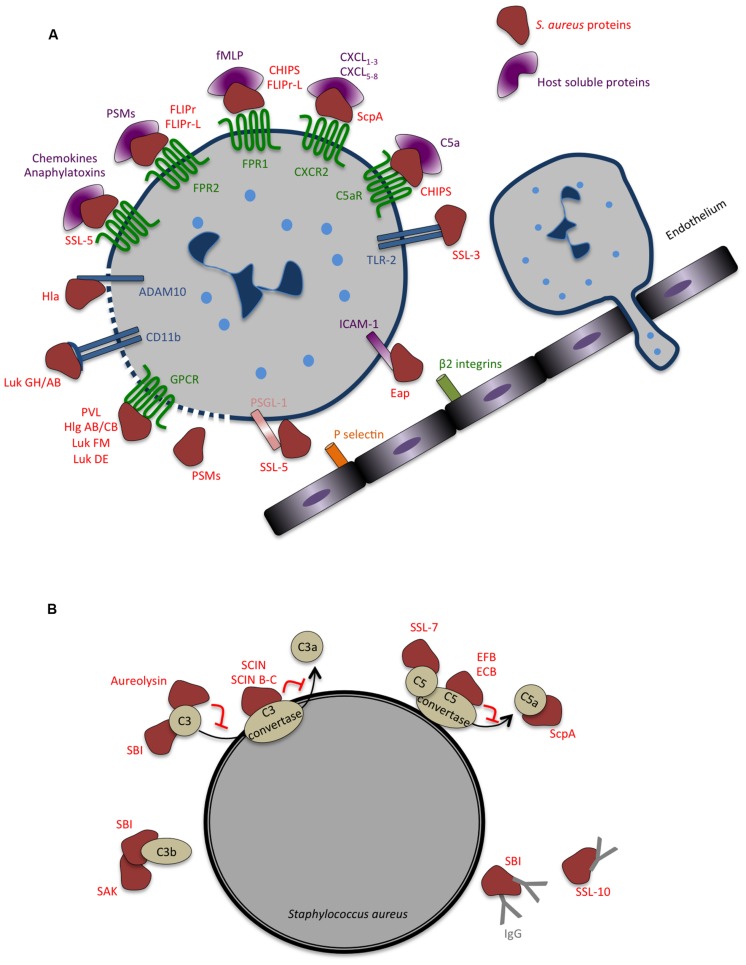
Bacterial toxins are virulence factors that manipulate host cell functions and take over the control of vital processes of living organisms to favor microbial infection. Some toxins directly target innate immune cells, thereby annihilating a major branch of the host immune response. In this review we will focus on bacterial toxins that act from the extracellular milieu and hinder the function of macrophages and neutrophils. In particular, we will concentrate on toxins from Gram-positive and Gram-negative bacteria that manipulate cell signaling or induce cell death by either imposing direct damage to the host cells cytoplasmic membrane or enzymatically modifying key eukaryotic targets. Outcomes regarding pathogen dissemination, host damage and disease progression will be discussed.

Keywords: bacterial exotoxin; immunomodulation; innate immune response; macrophage; neutrophil; phagocyte.


Figures

FIGURE 1
FIGURE 1
The concerted action of pertussis toxin (PT) and adenylate cyclase toxin (ACT) annihilates recruitment and function of innate immune cells. Following binding to a sialoglyprotein receptor, PT is endocytosed and retrogradely transported to the endoplasmic reticulum (ER). From the ER, the A subunit is delivered into the cytosol and travels to the plasma membrane, where it ADP-ribosylates the alpha-subunit of heterotrimeric G proteins, perturbing their regulatory functions and leading to an increase in the cAMP concentration that contributes to the early suppression of inflammatory cytokine production and inhibits the recruitment of immune cells to the site of infection. ACT binds with high affinity to CD11b/CD18 receptor [also known as complement receptor 3 (CR3) or macrophage-1 antigen (Mac-1)] present at the surface of macrophages, neutrophils, and dendritic cells. Upon binding, ACT integrates the membrane of target cell in two different conformations: a translocation precursor that relocalises at lipid raft domains from where the adenylate cyclase activity translocates directly to the cell cytoplasm; and a pore precursor that oligomerises and permeabilises the cells causing ion concentration imbalance. Binding of calmodulin stimulates the adenylate cyclase, leading to an increase in intracellular levels of cAMP. The activity of ACT inhibits complement-mediated phagocytosis, inhibits the production of pro-inflammatory cytokines and interferes with immune cell recruitment.
FIGURE 2
FIGURE 2
Anthrax toxins cooperatively disable host innate immune response. Host cell intoxication by anthrax toxins involves interaction of protective antigen (PA, 83 kDa) with two cellular receptors [tumor endothelium marker 8 (TEM8), also known as anthrax toxin receptor 1 (ANTXR1) and capillary morphogenesis protein 2 (CMG2) also known as anthrax toxin receptor 2 (ANTXR2)], which are expressed by different cell types, including macrophages and neutrophils. Upon binding to the cell surface receptor, PA83 is proteolytically processed at its N-terminus by a furin-like protease yielding the C-terminal fragment PA63 that oligomerises into a heptameric prepore able to bind edema factor (EF) and lethal factor (LF). The EF and/or LF-prepore-receptor complex undergoes receptor-mediated endocytosis and the acidic conditions in endosomes induce conversion of the prepore to a pore, allowing translocation of EF and LF into the cell cytosol to exert their cytotoxic effects. LT is a zinc-dependent metalloprotease that inhibits activation of neutrophils and macrophages, expression of inflammatory cytokines and cell motility by disrupting mitogen activated protein kinase kinases (MAPKKs)-regulated pathways. LT activity also promotes macrophage apoptosis by interfering with pro-survival MAPKK dependent pathways. ET is a calcium and calmodulin-dependent adenylate cyclase that increases intracellular cAMP concentration, leading to the suppression of the expression of inflammatory cytokines and cell chemotaxis through protein kinase A (PKA)-dependent pathways. The concerted action of LT and ET blocks the function of phagocytic cells.
FIGURE 3
FIGURE 3
AIP56 blocks innate immunity by inducing massive apoptosis of host macrophages and neutrophils. Upon encountering susceptible cells, apoptosis-inducing protein of 56 kDa (AIP56) binds to a still unidentified cell-surface receptor and undergoes clathrin-mediated endocytosis. Once in early endosomes, the toxin either follows the recycling pathway back to the extracellular medium or suffers low pH-induced translocation across the endosomal membrane into the cytosol to display its toxic activity. AIP56 is a zinc-dependent metalloprotease that cleaves the p65 subunit of nuclear factor-κB (NF-κB), an evolutionarily conserved transcription factor that regulates the expression of inflammatory and anti-apoptotic genes and plays a key role in host responses to microbial pathogen invasion. During infection, AIP56 disseminates systemically and its activity leads to depletion of macrophages and neutrophils by post-apoptotic secondary necrosis, thereby blocking the phagocytic defense of the host and contributing to the occurrence of tissue damage.
FIGURE 4
FIGURE 4
Strategies evolved by Staphylococcus aureus to counteract innate immune response. (A) Secreted bacterial factors that inhibit neutrophils extravasation, chemotaxis and activation. Neutrophil rolling is modulated by staphylococcal superantigen-like 5 (SSL5) that binds P-selectin glycoprotein ligand-1 (PSGL-1), blocking its interaction with P-selectin. The adhesion of neutrophils to the endothelium and consequent transmigration is inhibited by extracellular adherence protein (Eap), which binds to intercellular adhesion molecule 1 (ICAM-1). In addition to inhibiting PSGL-1, SSL5 inhibits neutrophil responses to chemokines and to anaphylatoxins, by binding to different chemokine receptors. Several staphylococcal molecules impair neutrophil chemotaxis and important co-signaling events during migration and phagocytosis: chemotaxis-inhibitor protein of S. aureus (CHIPS) binds and inhibits formyl peptide receptor 1 (FPR1) and C5a receptor (C5aR); formyl peptide receptor-like 1 inhibitor (FLIPr)-like inhibit FPR1; FLIPr and FLIPr-like inhibit FPR2; staphopain (ScpA) cleaves Chemokine (C–X–C Motif) Receptor 2 (CXCR2); staphylococcal SSL3 inhibits toll-like receptor 2 (TLR2)-mediated signaling, the bicomponent leukocidins Panton-Valentine leukocidin (PVL), gamma-hemolysin (Hlg) ABC, leukocidin (Luk) FM, Luk GH/AB, and Luk DE interact with chemoattractant receptors of the G-protein-coupled receptor (GPCR) family. Both Hla and Luk GH/AB induce cell lysis by binding ADAM metallopeptidase domain 10 (ADAM10) and CD11b, respectively. The cytolytic peptides phenol-soluble modulins (PSMs) have an amphipathic alpha-helical region that likely contributes to their lytic activity, presumably by membrane insertion and pore formation. (B) Secreted bacterial factors that inhibit opsonization and phagocytosis by neutrophils. The secreted metalloprotease aureolysin inhibits phagocytosis and killing of bacteria by neutrophils by cleaving C3. Staphylococcal complement inhibitor (SCIN), SCIN-B, and SCIN-C associate with and inhibits C3 convertase, thereby preventing the production of C3a, C3b, and further C5a and consequently interfering with complement activation. The extracellular fibrinogen binding protein (EFB) and the extracellular complement-binding protein (ECB) also inhibit complement activation by inactivating C5 convertase and staphylococcal SSL7 targets C5. Staphylococcal binder of immunoglobulin (SBI) affect both the function of complement and immunoglobulin binding, blocking the classical complement activation pathway, and associates with C3 inhibiting the alternative pathway. Staphylokinase (SAK) forms enzymatically active complexes with C3b blocking complement activation. Staphylococcal SSL10 binds IgG, affecting Fc receptor (FcR) recognition and complement activation.
FIGURE 5
FIGURE 5
Mycolactone inhibits the secretion of most cytokines, chemokines and other inflammatory mediators by macrophages. In eukaryotic cells, secretory proteins cross the ER membrane before being transported in vesicles to the Golgi complex and then to the plasma membrane. Mycolactone enters cells by passive diffusion through the plasma membrane and inhibits the production of inflammatory mediators by macrophages by blocking the translocation of nascent proteins into the ER. The proteins wrongly accumulated in the cytosol are then degraded by the proteasome.

References

    1. Abrami L., Lindsay M., Parton R. G., Leppla S. H., van der Goot F. G. (2004). Membrane insertion of anthrax protective antigen and cytoplasmic delivery of lethal factor occur at different stages of the endocytic pathway. J. Cell Biol. 166 645–651. 10.1083/jcb.200312072 - DOI - PMC - PubMed
    1. Abrami L., Liu S., Cosson P., Leppla S. H., van der Goot F. G. (2003). Anthrax toxin triggers endocytosis of its receptor via a lipid raft-mediated clathrin-dependent process. J. Cell. Biol. 160 321–328. 10.1083/jcb.200211018 - DOI - PMC - PubMed
    1. Adusumilli S., Mve-Obiang A., Sparer T., Meyers W., Hayman J., Small P. L. (2005). Mycobacterium ulcerans toxic macrolide, mycolactone modulates the host immune response and cellular location of M. ulcerans in vitro and in vivo. Cell. Microbiol. 7 1295–1304. 10.1111/j.1462-5822.2005.00557.x - DOI - PubMed
    1. Agrawal A., Lingappa J., Leppla S. H., Agrawal S., Jabbar A., Quinn C., et al. (2003). Impairment of dendritic cells and adaptive immunity by anthrax lethal toxin. Nature 424 329–334. 10.1038/nature01794 - DOI - PubMed
    1. Ahmad J. N., Cerny O., Linhartova I., Masin J., Osicka R., Sebo P. (2015). cAMP signaling of Bordetella adenylate cyclase toxin through the SHP-1 phosphatase activates the BimEL-Bax pro-apoptotic cascade in phagocytes. Cell Microbiol. 10.1111/cmi.12519 [Epub ahead of print]. - DOI - PubMed

LinkOut - more resources