

Clinicopathological features of dermatofibrosarcoma protuberans
- PMID: 26870263
- PMCID: PMC4726970
- DOI: 10.3892/ol.2015.3966
Clinicopathological features of dermatofibrosarcoma protuberans
Abstract
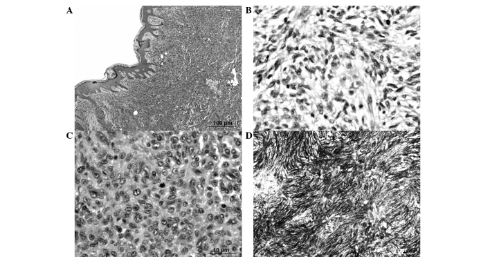
Dermatofibrosarcoma protuberans (DFSP) is a superficial cutaneous tumor of low malignant potential characterized by a high rate of local recurrence. The histopathological appearance shows uniform spindle neoplastic cells arranged in a predominantly storiform pattern, typically with positive staining for cluster of differentiation (CD)34 and vimentin on immunohistochemistry. A minority of cases of DFSP have areas of sarcomatous transformation. Wide surgical excision is the cornerstone of treatment for DFSP. The objective of the present study was to determine the clinicopathological features of DFSP. Pathological records were searched for cases of DFSP in the database of the Department of Pathology, Faculty of Medicine Ramathibodi Hospital (Mahidol University, Bangkok, Thailand) between 1994 and 2013. The results showed 68 cases with DFSP. The mean age at diagnosis was 40 years (range, 3-86 years). Among this group of patients, 26 cases (38.2%) experienced local recurrence and 6 (8.8%) exhibited sarcomatous transformation of DFSP. The factors that predict the recurrence of DFSP are an incorrect first pathological diagnosis and an inadequate surgical margin. The factors that predict the sarcomatous transformation of DFSP are a larger tumor size and an incorrect first pathological diagnosis. In patients who have tumors with spindle cells arranged in a storiform pattern, CD34 immunohistochemical staining provides the definitive diagnosis. Exact histopathological categorization is important to select the appropriate treatment and predict the clinical outcome.
Keywords: aggressive cutaneous tumor; dermatofibrosarcoma protuberans; recurrence; sarcomatous transformation.
Figures
References
-
- Mentzel T, Peeutour F, Lazar A, Coindre JM. Dermatofibrosarcoma protuberans. World Health Organization (WHO) Classification of Tumours of Soft tissue and Bone. In: Fletcher CDM, Bridge JA, Hogendoorn P, Martens F, editors. Pathology and Genetics. 4th. Vol. 5. Lyon: IARC Press; 2013. pp. 77–79.
-
- Weyers W, Mentzel T, Kasper RC, Tosti A, Iorizzo M, Zelger B, Caputo R. In: World Health Organization Classification of Tumours. Pathology and Genetics Skin Tumours. In: Le Boit PE, Burg G, Weedon D, Sarasin A, editors. Fibrous, fibrohistiocytic and histiocytic tumours. Lyon: IARC Press; 2002. pp. 259–261.
-
- Kempson RL, Fletcher CDM, Evans HL, Hendrickson MR, Sibley RK. Armed Forces Institute of Pathology. Washington, DC: 2001. Atlas of Tumor Pathology. Tumors of Soft Tissues. 3rd series. Fascicle 30; pp. 138–148.
-
- Goldblum JR, Folpe AL, Weiss SW, editors. Enzinger and Weisss soft tissue tumors. 6th. Philadelphia, PA: Elsevier Saunders; 2013. Fibrohistocytic tumors of intermediate malignancy; pp. 387–400.
-
- Darier J, Ferrand M. Dermatofibromes progressifs et récidivants ou fibrosarcomes de la peau. Ann Dermatol Syphiligr (Paris) 1924;5:545–562.
LinkOut - more resources
Full Text Sources
Other Literature Sources