. 2015 Dec 1;8(12):3912-3921.
doi: 10.1007/s12274-015-0891-y.

An ultra-sensitive dual-mode imaging system using metal-enhanced fluorescence in solid phantoms

Affiliations

An ultra-sensitive dual-mode imaging system using metal-enhanced fluorescence in solid phantoms

Eran A Barnoy et al. Nano Res. .

Abstract

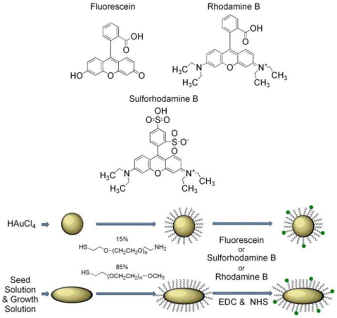
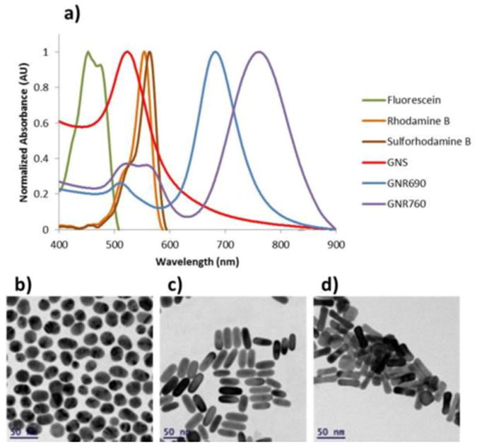
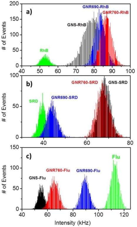
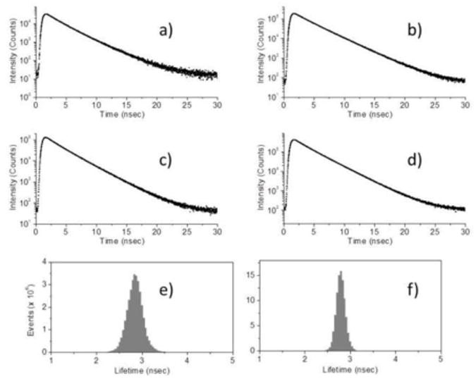
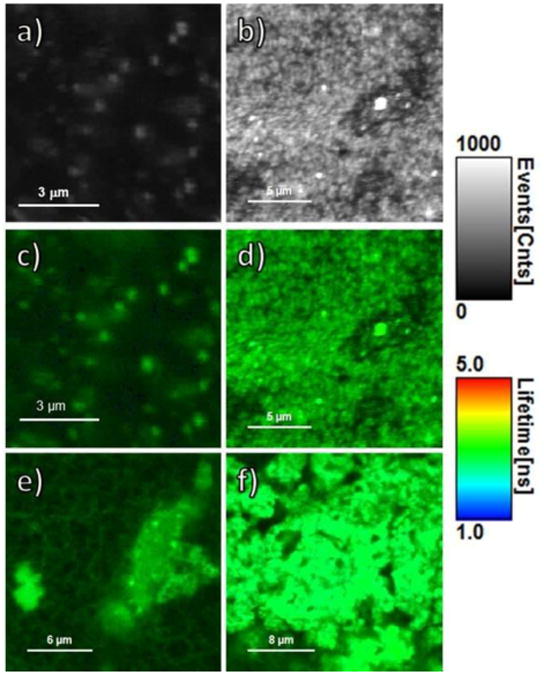
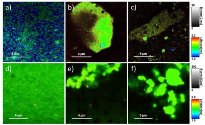
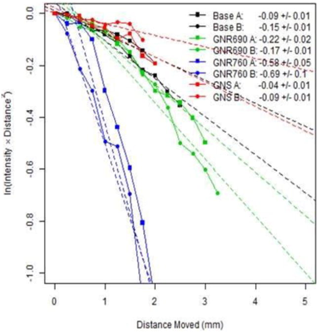
In this study we developed a highly sensitive dual modal imaging system designed for gold nanoparticles (GNPs) conjugated to various fluorophores in solid phantoms. The system consists of fluorescence lifetime imaging microscopy (FLIM) for surface imaging, diffusion reflection (DR) for deep tissue imaging (up to 1cm), and metal enhanced fluorescence (MEF). We detected quenching in fluorescent intensity (FI) for the conjugation of gold nanospheres (GNS) as well as gold nanorods (GNRs) to Fluorescein, which has an excitation peak at a wavelength shorter than the surface plasmon resonance (SPR) of both types of GNPs, and enhanced FI in conjugation to Rhodamine B and Sulforhodamine B, both with excitation peaks in the GNPs' SPR. The enhanced FI was detected in solution as well as in solid phantoms from FLIM measurements. DR measurements detected GNR presence within the solid phantoms by recording dropped rates of light scattering using wavelengths corresponding to the GNRs' absorption. With the inclusion of MEF, this promising dual modal imaging technique enables efficient and sensitive molecular and functional imaging.

Keywords: Gold nanoparticles; biomolecular imaging; diffusion reflection; fluorescence lifetime imaging; metal enhanced fluorescence; noninvasive detection.


Figures

Figure 1
Figure 1
Schematic representation of gold nanospheres (GNSs) and gold nanorods (GNRs) preparation and subsequent conjugation to fluorophores.
Figure 2
Figure 2
Particle characteristics. (a) Normalized absorption spectra of GNS, GNR690, and GNR760 alongside that of Fluorescein, Rhodamine B, and Sulforhodamine B, and transmission electron microscope (TEM) images of synthesized (b) GNS, (c) GNR690, and (d) GNR760.
Figure 3
Figure 3
Fluorescence Intensity (FI) enhancement and quenching due to GNP conjugation in solution. FI is depicted in photon counts/msec, or kHz. (a) Count rate histograms of GNS, GNR690 and GNR760 conjugated to Rhodamine B (RhB). FI of RhB solution without GNPs is also included in the panel for comparison. (b) Count rate histograms of GNS, GNR690 and GNR760 conjugated to Sulforhodamine B (SRD). FI of SRD is included in the panel for comparison. (c) Count rate histograms of GNS, GNR690 and GNR760 conjugated to Fluorescein (Flu). FI of Flu is included in the panel for comparison. All solutions contained a fluorophore concentration of 1μM. All measurements for a given fluorophore were obtained under the same conditions, same set-up, and same excitation power.
Figure 4
Figure 4
Representative fluorescent lifetime (FLT) curves obtained from FLT imaging microscopy (FLIM) for phantoms with RhB base containing (a) RhB only, (b) GNS-RhB, (c) GNR690-RhB, and (d) GNR780-RhB. Representative FLT histograms for the same phantoms containing (e) GNR690-RhB and (f) GNR780-RhB. All curves were obtained under the same conditions, same set-up, and same excitation power.
Figure 5
Figure 5
FLIM images of phantoms with RhB base. Depicting only FI (shown in counts of fluorescence events), the images show phantoms containing (a) RhB only and (b) GNS-RhB. Combining FI (shown as brightness) and FLT (shown as color), the images show phantoms containing (c) RhB only, (d) GNS-RhB, (e) GNR690-RhB and (f) GNR760-RhB. For all 6 images, the gray, brightness scale bar represents FI in counts/msec. The color scale bar displays the FLT range in nsec. All images were obtained under the same conditions, same set-up, and same excitation power.
Figure 6
Figure 6
FLIM images of phantoms containing (a) GNS-SRD, (b) GNR690-SRD, (c) GNR760-SRD, (d) GNS-Flu, (e) GNR690-Flu and (f) GNR760-Flu. Each gray scale bar applies to all images in a given row, and represents FI in counts/msec. Each color scale bar applies to all images in a given row, and displays the FLT range in nsec. All images for a given fluorophore were obtained under the same conditions, same set-up, and same excitation power.
Figure 7
Figure 7
Diffusion Reflection (DR) measurements of scattered light intensity as a function of distance from 4 different phantoms, each denoted by a different color: a water-only base (black), GNR690 (green), GNR760 (blue), and GNS (red). Each phantom was measured 2 times, as denoted by the marker shapes. The light source had wavelength 780nm, and the concentration of the particles in the phantoms was set so that they would contain a fluorophore concentration of 5μM. The legend depicts the slope ± standard error to the slope of a fitted line to the corresponding curve. As explained in Eq. (2), this slope is an indication of the spectral properties of the sample.

Similar articles

Cited by

References

    1. Cao YC, Jin R, Mirkin CA. Nanoparticles with Raman spectroscopic fingerprints for DNA and RNA detection. Science. 2002;297:1536–1540. - PubMed
    1. Rosi NL, Mirkin CA. Nanostructures in biodiagnostics. Chem Rev. 2005;105:1547–1562. - PubMed
    1. Nie S, Xing Y, Kim GJ, Simons JW. Nanotechnology applications in cancer. Annu Rev Biomed Eng. 2007;9:257–288. - PubMed
    1. Eghtedari M, Liopo AV, Copland JA, Oraevsky AA, Motamedi M. Engineering of hetero-functional gold nanorods for the in vivo molecular targeting of breast cancer cells. Nano Lett. 2009;9:287–291. - PMC - PubMed
    1. Von Maltzahn G, Park J, Agrawal A, Bandaru NK, Das SK, Sailor MJ, Bhatia SN. Computationally guided photothermal tumor therapy using long-circulating gold nanorod antennas. Cancer Res. 2009;69:3892–3900. - PMC - PubMed

LinkOut - more resources