. 2015 Oct 3;2(11):1785-98.
doi: 10.1016/j.ebiom.2015.09.053. eCollection 2015 Nov.

Stress Granules Modulate SYK to Cause Microglial Cell Dysfunction in Alzheimer's Disease

Affiliations

Stress Granules Modulate SYK to Cause Microglial Cell Dysfunction in Alzheimer's Disease

Soumitra Ghosh et al. EBioMedicine. .

Erratum in

Abstract

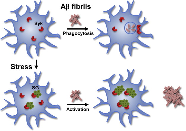
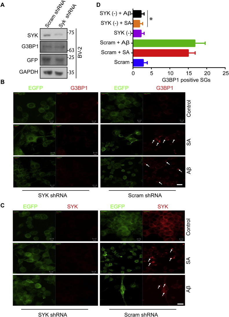
Microglial cells in the brains of Alzheimer's patients are known to be recruited to amyloid-beta (Aβ) plaques where they exhibit an activated phenotype, but are defective for plaque removal by phagocytosis. In this study, we show that microglia stressed by exposure to sodium arsenite or Aβ(1-42) peptides or fibrils form extensive stress granules (SGs) to which the tyrosine kinase, SYK, is recruited. SYK enhances the formation of SGs, is active within the resulting SGs and stimulates the production of reactive oxygen and nitrogen species that are toxic to neuronal cells. This sequestration of SYK inhibits the ability of microglial cells to phagocytose Escherichia coli or Aβ fibrils. We find that aged microglial cells are more susceptible to the formation of SGs; and SGs containing SYK and phosphotyrosine are prevalent in the brains of patients with severe Alzheimer's disease. Phagocytic activity can be restored to stressed microglial cells by treatment with IgG, suggesting a mechanism to explain the therapeutic efficacy of intravenous IgG. These studies describe a mechanism by which stress, including exposure to Aβ, compromises the function of microglial cells in Alzheimer's disease and suggest approaches to restore activity to dysfunctional microglial cells.

Keywords: AD, Alzheimer's disease; Alzheimer's disease; Amyloid-beta; Aβ, amyloid-beta; IgG, immunoglobulin G; MG, microglial cells; Microglial cells; Neurodegenerative disease; SYK tyrosine kinase; Stress granules.


Figures

None
Graphical abstract
Fig. 1
Fig. 1
SYK associates with SGs in MG. (A) Lysates of BV-2, N9 and DG75 cells were examined by Western blotting using antibodies against SYK and GAPDH. (B) BV-2 and N9 cells were treated without (control) or with SA (A) for 24 h, fixed and stained for SYK (green) and G3BP1 (red). Examples of SGs are indicated by the arrows. Bar = 10 μm. (C) The area occupied by SYK and G3BP1 positive SGs in N9 cells was quantified by Image J analysis of 5 random frames from 3 independent experiments. Results represent means ± SEM. *P = 0.001. (D) BV-2 and N9 cells were treated without (control) or with soluble Aβ for 24 h, fixed and stained for SYK (green) and G3BP1 (red). Examples of SGs are indicated by the arrows. Bar = 10 μm. (E) BV-2 cells were treated with soluble Aβ(42–1) for 24 h, fixed and stained for SYK (green) and G3BP1 (red). Only merged images are shown.
Fig. 2
Fig. 2
Characterization of SGs in MG. (A and B) N9 cells were treated without (control) or with SA or soluble Aβ for 24 h, fixed and stained for SYK, pEIF2α, PABP1, TDP-43, TIA-1 and/or G3BP1 as indicated. Only merged images are displayed. Examples of SGs are indicated by the arrows. Bar = 10 μm. (C) N9 cells were treated without (−) or with (+) SA or soluble Aβ for 24 h and then separated into detergent soluble and insoluble fractions, which were analyzed by Western blotting for SYK, G3BP1, TIA-1 and pEIF2α. Data shown are representative of three separate experiments.
Fig. 3
Fig. 3
SYK associates with SGs in MG from aged mice. (A and B) MG isolated from the brains of 20 month old wild-type (A) or Syk+/− (B) mice were treated without (control) or with SA or soluble Aβ for 24 h, fixed and co-stained for IBA1 and SYK or IBA1 and G3BP1. Examples of SGs are indicated by the arrows. Bar = 5 μm. (C) Quantitative comparison of SG formation in MG from young (1 m) and old (20 m), wild-type (Wt) and Syk haploinsufficient (Het) mice. **P = 0.001.
Fig. 4
Fig. 4
SYK is active in SGs and promotes their formation. (A) N9 cells were treated without (control) or with SA or soluble Aβ for 24 h, fixed and stained for SYK (green) and phosphotyrosine (pTYR, red). Examples of SGs are indicated by the arrows. Bar = 10 μm. (B) Phosphotyrosine-containing proteins were immunoprecipitated from lysates of N9 cells treated without (control) or with SA or soluble Aβ for 24 h. Immune complexes were examined by Western blotting with antibodies against SYK or phosphotyrosine (pTYR). SYK in the original lysates was detected by Western blotting (input). Beads lacking anti-phosphotyrosine were used as a control (mock). (C) Lysates from N9 cells treated without (control) or with SA or soluble Aβ for 24 h were analyzed by Western blotting using antibodies against SYK phosphorylated on Y519 and Y520 (p-SYK), SYK or GAPDH. (D) N9 cells were treated without (control) or with SA or soluble Aβ for 24 h in the presence or absence of R406 or PRT318 (PRT) (500 nM each), fixed and stained for SYK (green) and G3BP1 (red). Examples of SGs are indicated by the arrows. Bar = 10 μm. (E) The number of SGs formed per cell from (D). Results represent means ± SEM from 3 independent experiments. *P = 0.0001.
Fig. 5
Fig. 5
Reduced SYK inhibits SG formation in MG. (A) BV-2 cells expressing EGFP and scrambled shRNA or SYK shRNA were examined by Western blotting with antibodies against SYK, G3BP1, GFP and GAPDH. (B and C) BV-2 cells infected with a lentivirus expressing EGFP and either a SYK or scrambled shRNA were treated without (control) or with SA or soluble Aβ for 24 h, fixed and stained for G3BP1 (B) or SYK (C) (both in red). Examples of SGs are indicated by the arrows. Bar = 10 μm. (D) Quantification of G3BP1 positive SGs present within 10 random 25 × frames for cells expressing SYK shRNA (SYK (−)) or scrambled shRNA (Scram). *P = 0.001.
Fig. 6
Fig. 6
Chronic stress leads to large SGs and persistent ROS and RNS production. (A) N9 cells were treated without (control) or with SA or soluble Aβ for 5 days. Where indicated, cells were cultured in the absence of SA or Aβ for an additional 48 h. Cells were fixed and stained for SYK (green) and G3BP1 (red). Examples of SGs are indicated by the arrows. Bar = 10 μm. (B-D) N9 cells were treated without (Control) or with SA or Aβ for 5 days in the presence or absence of R406 or PRT318 (500 nM each). Cells were incubated with carboxy-DCFDA (10 μM) for 1 h and examined using a fluorescence plate reader (B). Media was collected and analyzed for ROS using a chemiluminescence assay (C) or nitrite levels using a fluorescence assay (D). Data represent means ± SEM for 3 independent experiments. *P = 0.001, **P = 0.0001. (E) N9 cells treated without (control) or with SA or Aβ for 5 days in the presence or absence of PRT318 (500 nM) were incubated with D123 for 1 h and green fluorescence was observed by confocal microscopy. Hoechst dye (blue) was used to stain the nucleus. Bar = 10 μm. (F) N9 cells treated for 5 days with SA or Aβ were washed thoroughly and then co-cultured with HT22 for another 48 h separated by a transwell insert. HT22 cells were incubated with FITC Annexin V (green) for 30 min, fixed and imaged by confocal microscopy. Hoechst dye (blue) was used to stain the nucleus. Bar = 10 μm. (G) Lysates from the cells in (D) were analyzed by Western blotting for NOS2 and GAPDH.
Fig. 7
Fig. 7
SYK inhibitors or chronic stress block MG phagocytosis. (A) BV-2 cells were treated without (control) or with SA or soluble Aβ for 5 days in the presence or absence of R406 or PRT381 (500 nM each) and examined for the uptake of pHRodo particles (red). Cells were fixed and examined by confocal microscopy. Hoechst dye (blue) was used to stain the nucleus. Bar = 10 μm. (B) Quantification of (A) showing change in mean fluorescence measured by Image J from ten random frames in three independent experiments. *P = 0.001 as compared to control cells, **P = 0.0001 as compared to control cells. (C) N9 cells treated without (control) or with SA or soluble Aβ for 5 days were incubated with FITC-labeled Aβ fibrils for 12 h, washed, fixed and examined by confocal microscopy. Hoechst dye (blue) was used to stain the nucleus.
Fig. 8
Fig. 8
SYK, G3BP1 and phosphotyrosine-positive SGs are found in AD brains. (A–C) Samples from the cortex region of human brain from patients with no (normal), mild, moderate (mod) and severe AD were immunostained for IBA1, SYK, G3BP1 and/or phosphotyrosine (pTYR) as indicated. Bar = 5 μm.
Fig. 9
Fig. 9
Restoration of phagocytic activity with IgG. (A and B) N9 cells were treated without (control) or with SA or soluble Aβ for 120 h in the presence or absence of R406 or PRT318 (500 nM each). At 116 h, to one set of control, SA- and Aβ-treated cells was added anti-β1-integrin (β-Int), mixed rabbit IgG (IgG) or anti-CD32 for the final 4 h. Cells were then incubated with pHRhodo particles (red) to measure phagocytosis (A) or fixed and stained with antibodies against SYK (B). Hoechst dye (blue) was used to stain the nucleus (A). Bar = 10 μm.

Comment in

  • Syk and Yea Shall Find.
    Wolozin B, Ikezu T. Wolozin B, et al. EBioMedicine. 2015 Nov 10;2(11):1590-1. doi: 10.1016/j.ebiom.2015.11.012. eCollection 2015 Nov. EBioMedicine. 2015. PMID: 26870779 Free PMC article. No abstract available.

References

    1. Anderson P., Kedersha N. Stress granules: the Tao of RNA triage. Trends Biochem. Sci. 2008;33:141–150. - PubMed
    1. Angata T., Kerr S.C., Greaves D.R., Varki N.M., Crocker P.R., Varki A. Cloning and characterization of human siglec-11: a recently evolved signaling molecule that can interact with SHP-1 and SHP-2 and is expressed by tissue macrophages, including brain microglia. J. Biol. Chem. 2002;277:24466–24474. - PubMed
    1. Ash P.E., Vanderweyde T.E., Youmans K.L., Apicco D.J., Wolozin B. Pathological stress granules in Alzheimer's disease. Brain Res. 2014;1584:52–58. - PMC - PubMed
    1. Bamberger M.E., Harris M.E., McDonald D.R., Husemann J., Landreth G.E. A cell surface receptor complex for fibrillar beta-amyloid mediates microglial activation. J. Neurosci. 2003;23:2665–2674. - PMC - PubMed
    1. Barcia C., Ross C.M., Annese V., Gómez A., Ros-Bernal F., Aguado-Llera D., Martinez-Pagán E., de Pablos V., Fernandez-Villalba E., Herrero M.T. IFN-γ signaling, with the synergistic contribution of TNF-α, mediates cell specific microglial and astroglial activation in experimental models of Parkinson's disease. Cell Death Dis. 2012;3 - PMC - PubMed

Publication types

MeSH terms

LinkOut - more resources