. 2015 Oct 19;2(11):1806-13.
doi: 10.1016/j.ebiom.2015.10.017. eCollection 2015 Nov.

The Sigma-2 Receptor and Progesterone Receptor Membrane Component 1 are Different Binding Sites Derived From Independent Genes

Affiliations

The Sigma-2 Receptor and Progesterone Receptor Membrane Component 1 are Different Binding Sites Derived From Independent Genes

Uyen B Chu et al. EBioMedicine. .

Abstract

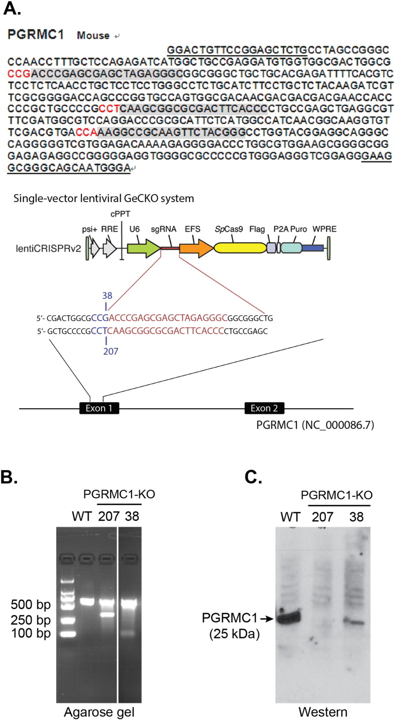
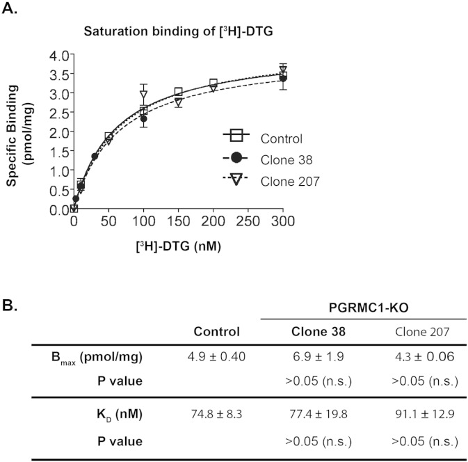
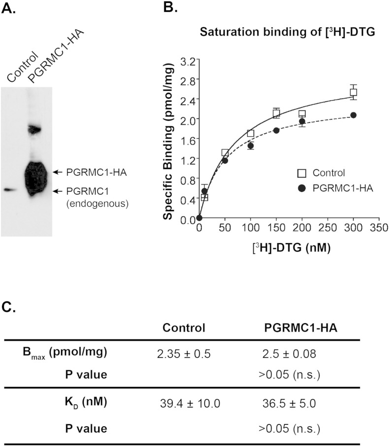
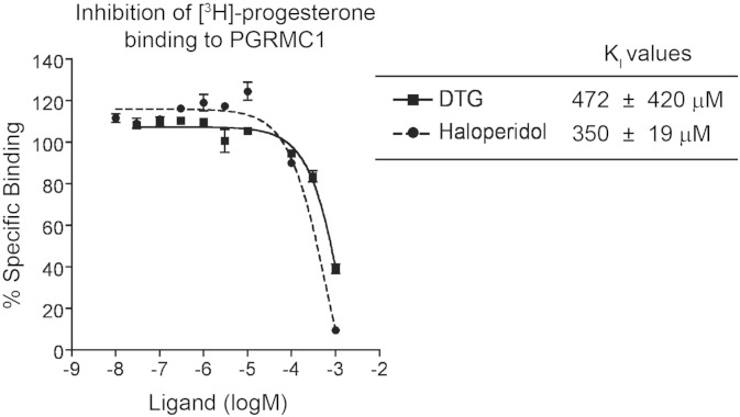
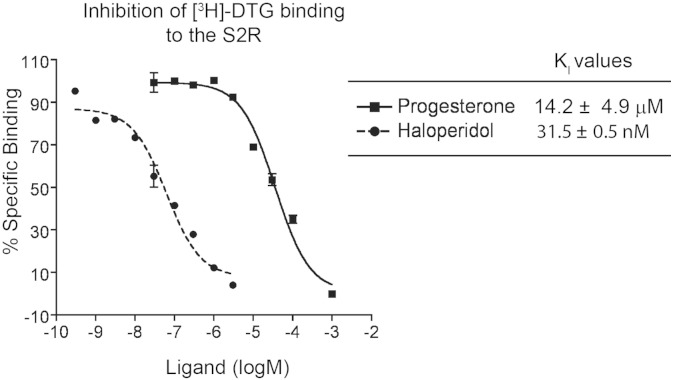
The sigma-2 receptor (S2R) is a potential therapeutic target for cancer and neuronal diseases. However, the identity of the S2R has remained a matter of debate. Historically, the S2R has been defined as (1) a binding site with high affinity to 1,3-di-o-tolylguanidine (DTG) and haloperidol but not to the selective sigma-1 receptor ligand (+)-pentazocine, and (2) a protein of 18-21 kDa, as shown by specific photolabeling with [(3)H]-Azido-DTG and [(125)I]-iodoazido-fenpropimorph ([(125)I]-IAF). Recently, the progesterone receptor membrane component 1 (PGRMC1), a 25 kDa protein, was reported to be the S2R (Nature Communications, 2011, 2:380). To confirm this identification, we created PGRMC1 knockout NSC34 cell lines using the CRISPR/Cas9 technology. We found that in NSC34 cells devoid of or overexpressing PGRMC1, the maximum [(3)H]-DTG binding to the S2R (Bmax) as well as the DTG-protectable [(125)I]-IAF photolabeling of the S2R were similar to those of wild-type control cells. Furthermore, the affinities of DTG and haloperidol for PGRMC1 (KI = 472 μM and 350 μM, respectively), as determined in competition with [(3)H]-progesterone, were more than 3 orders of magnitude lower than those reported for the S2R (20-80 nM). These results clarify that PGRMC1 and the S2R are distinct binding sites expressed by different genes.

Keywords: Clustered regularly interspaced short palindromic repeats (CRISPR)/Cas9 knockout; Progesterone receptor membrane component-1 (PGRMC1); Sigma-2 receptor (S2R); [125I]-Iodoazido-fenpropimorph ([125I]-IAF); [3H]-1,3-Di-o-tolylguanidine ([3H]-DTG).


Figures

Fig. 1
Fig. 1
PGRMC1 knockout using a CRISPR/Cas9 approach. A. Schematic of the PGRMC1 sgRNA/Cas9-expressing lentiviral constructs for knocking out PGRMC1. Gray areas show the three candidate sgRNA sequences in the exon-1 of the PGRMC1 gene, of which two were used for the CRISPR/Cas9 constructs. B. A representative DNA gel of control and PGRMC1 knockout (clones 38 and 207) NSC34 cells verifying the Cas9 cleavage of the genomic DNA. Control refers to the NSC34 cells transfected with the control vector expressing Cas9 but not an sgRNA. C. Western blotting detection of PGRMC1 in control and PGRMC1 knockout (clones 38 and 207) NSC34 cells.
Fig. 2
Fig. 2
[125I]-IAF photolabeling of the S2R is protected by S2R-specific ligands but not affected by PGRMC1 knockout. A. [125I]-IAF photolabeling of the S2R in PC12 membranes is protectable by CM compounds. [125I]-IAF photolabeling of both S1R and S2R was protected by 20 μM DTG (D) while CM compounds (CM 353, CM 398, CM 775, and CM 777, see Fig. S1 for affinities to sigma receptors) (Matsumoto et al., 2014) selectively blocked the labeling of the S2R but not the S1R. Two concentrations (1 and 10 μM) were used for CM compounds. B. [125I]-IAF photolabeling of the S2R in membranes prepared from control and PGRMC1 knockout (clones 38 and 207) NSC34 cells. 5 μM (+)-pentazocine (P) protected against [125I]-IAF photolabeling of the S1R while 20 μM DTG (D) protected against photolabeling of both the S1R and the S2R. Note that the background photolabeled bands were not protected by these specific sigma ligands.
Fig. 3
Fig. 3
Eliminating PGRMC1 protein does not alter [3H]-DTG binding to the S2R in cell membranes. A. A representative of [3H]-DTG saturation binding in membranes prepared from control and PGRMC1 knockout (clones 38 and 207) NSC34 cells, (+)-pentazocine (100 nM) was included to mask [3H]-DTG binding to the S1R such that [3H]-DTG would be bound only to the S2R and measured as specific S2R binding. Nonspecific binding was measured (by adding haloperidol) and subtracted. Control refers to the NSC34 cells transfected with the control vector for the expression of Cas9 but not an sgRNA. B. Statistics. Maximum binding (Bmax) and equilibrium dissociation constants (KD) for [3H]-DTG were calculated using a Prizm software and reported as mean ± SEM of three separate experiments each performed in triplicates. Not significant (n.s.).
Fig. 4
Fig. 4
PGRMC1 overexpression does not change [3H]-DTG binding to the S2R in cell membranes. A. A representative Western blot detecting PGRMC1 in membranes prepared from control and PGRMC1-3 × HA overexpressing NSC34 cells. Control refers to the NSC34 cells transfected with the same expression vector but without the PGRMC1 gene. B. A representative experiment of [3H]-DTG saturation binding (as described in Fig. 3A) in membranes prepared from control and PGRMC1-3 × HA overexpressing NSC34 cells. C. Statistics. Maximum binding (Bmax) and equilibrium dissociation constants (KD) for [3H]-DTG were calculated using a Prizm software and reported as mean ± SEM of three separate experiments each performed in triplicates. Not significant (n.s.).
Fig. 5
Fig. 5
Assessment of DTG and haloperidol binding affinities for PGRMC1. Experiments were performed with rat liver membranes. Shown on the left is a representative inhibition curve of [3H]-progesterone binding (at 30 nM) to PGRMC1 in the presence of increasing concentrations of non-radioactive DTG or haloperidol. Nonspecific binding was measured by the addition of 10 μM of nonradioactive progesterone and subtracted, as previously described by Peluso et al. (2008). Inhibition constants (KI) are reported as mean ± SEM from three separate experiments each performed in triplicates for DTG and two separate experiments each performed in triplicates for haloperidol.
Fig. 6
Fig. 6
Assessment of progesterone and haloperidol binding affinities for the S2R. Experiments were performed with rat liver membranes. Shown on the left is a representative inhibition curve of [3H]-DTG binding (at 60 nM) to the S2R in the presence of increasing concentrations of non-radioactive progesterone or haloperidol. Nonspecific binding was measured by the addition of 10 μM of nonradioactive haloperidol and subtracted. Inhibition constants (KI) are reported as mean ± SEM from 3 separate experiments each performed in triplicates.

Similar articles

Cited by

References

    1. Abate C., Niso M., Infantino V., Menga A., Berardi F. Elements in support of the 'non-identity' of the PGRMC1 protein with the sigma2 receptor. Eur. J. Pharmacol. 2015;758:16–23. - PubMed
    1. Bali N., Arimoto J.M., Morgan T.E., Finch C.E. Progesterone antagonism of neurite outgrowth depends on microglial activation via Pgrmc1/S2R. Endocrinology. 2013;154:2468–2480. - PMC - PubMed
    1. Bowen W.D., Hellewell S.B., Mcgarry K.A. Evidence for a multi-site model of the rat brain sigma receptor. Eur. J. Pharmacol. 1989;163:309–318. - PubMed
    1. Cassano G., Gasparre G., Contino M., Niso M., Berardi F., Perrone R., Colabufo N.A. The sigma-2 receptor agonist PB28 inhibits calcium release from the endoplasmic reticulum of SK-N-SH neuroblastoma cells. Cell Calcium. 2006;40:23–28. - PubMed
    1. Chu U.B., M.T A., Chu M.-L., Yang H., Mesangeau C., McCurdy C.R., Guo L.-W., Ruoho A.E. Experimental Biology Meeting. 2015. The 18 kDa sigma-2 receptor and PGRMC1 are derived from separate genes. (LB511)

Publication types

MeSH terms