

Arterial Blood, Rather Than Venous Blood, is a Better Source for Circulating Melanoma Cells
- PMID: 26870807
- PMCID: PMC4740300
- DOI: 10.1016/j.ebiom.2015.09.019
Arterial Blood, Rather Than Venous Blood, is a Better Source for Circulating Melanoma Cells
Abstract
Background: CTCs provide prognostic information and their application is under investigation in multiple tumor types. Of the multiple variables inherent in any such process, none is more important to outcome than the appropriateness of the sample source. To address this question, we investigated CTCs in paired peripheral venous and arterial blood specimens obtained from stage IV uveal melanoma patients.
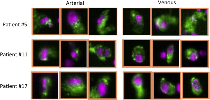
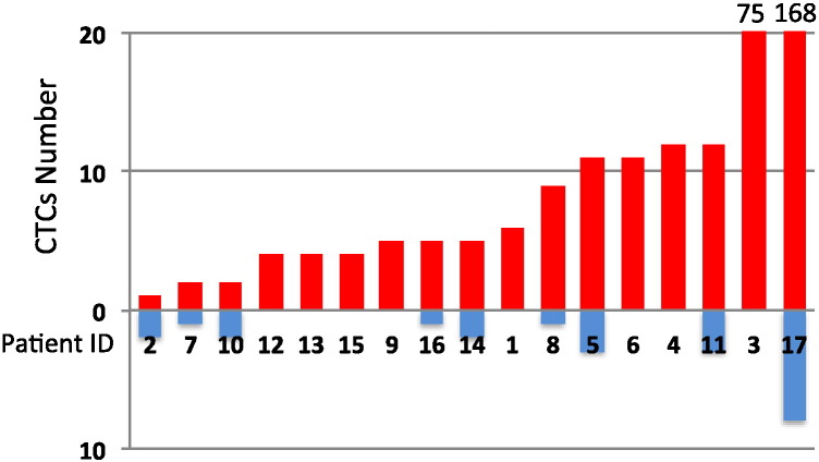
Methods: Blood specimens were obtained from both common femoral arteries and antecubital veins in 17 uveal melanoma patients with multiple hepatic metastases for CTC measurements.
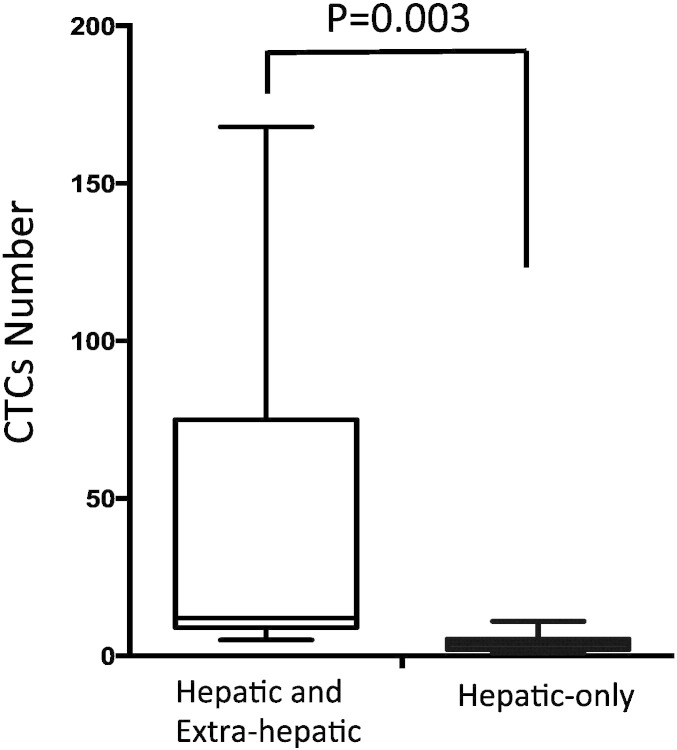
Finding: CTCs were detectable with greater frequency (100%) and in larger numbers (median 5, range 1 to 168) in all arterial blood specimens than in venous samples (52.9%; median 1, range 0 to 8). Patients with hepatic as well as extra-hepatic metastasis showed higher number of arterial CTCs, compared to patients with liver-only metastasis (p = 0.003). There was no significant association between the number of arterial CTCs and the tumor burden within the liver in patients who had liver-only metastases.
Interpretation: Our data indicate that arterial blood specimens might be a better source of circulating uveal melanoma cells. Although less conveniently processed, perhaps arterial blood should be evaluated as sample source for measurement of CTCs.
Keywords: AKTi, AKT inhibitor; Ab, antibody; Arterial venous; BCNU, bischlorethylnitrosourea; CTC count; Circulating tumor cells; DEBDOX, drug-eluting beads with doxorubicin; EDTA, ethylenediaminetetraacetic acid; HMW-MAA, high molecular weight melanoma associated antigen; Hepatic metastasis; Ipi, ipilimumab; LN, lymph node; MEKi, MEK inhibitor; METi, MET inhibitor;; Peripheral venous; TACE, transarterial chemoembolization; Uveal melanoma; VPA, valproic acid; XRT, radiation therapy.
Figures

Comment in
-
Arterial or Venous: Where Are the Circulating Tumor Cells?EBioMedicine. 2015 Sep 16;2(11):1596-7. doi: 10.1016/j.ebiom.2015.09.029. eCollection 2015 Nov. EBioMedicine. 2015. PMID: 26870782 Free PMC article. No abstract available.
References
-
- All-Ericsson C., Girnita L., Seregard S., Bartolazzi A., Jager M.J., Larsson O. Insulin-like growth factor-1 receptor in uveal melanoma: a predictor for metastatic disease and a potential therapeutic target. Invest. Ophthalmol. Vis. Sci. 2002;43(1):1–8. - PubMed
-
- Bidard F.C., Madic J., Mariani P., Piperno-Neumann S., Rampanou A., Servois V., Cassoux N., Desjardins L., Milder M., Vaucher I. Detection rate and prognostic value of circulating tumor cells and circulating tumor DNA in metastatic uveal melanoma. Int. J. Cancer. 2014;134(5):1207–1213. - PubMed
-
- Budd G.T., Cristofanilli M., Ellis M.J., Stopeck A., Borden E., Miller M.C., Matera J., Repollet M., Doyle G.V., Terstappen L.W. Circulating tumor cells versus imaging–predicting overall survival in metastatic breast cancer. Clin. Cancer Res. 2006;12(21):6403–6409. - PubMed
-
- Burger J.A., Kipps T.J. CXCR4: a key receptor in the crosstalk between tumor cells and their microenvironment. Blood. 2006;107(5):1761–1767. - PubMed
Publication types
MeSH terms
Grants and funding
LinkOut - more resources
Full Text Sources
Other Literature Sources
Medical
Miscellaneous
