. 2016 Apr 19;7(16):21428-40.
doi: 10.18632/oncotarget.7258.

RHAMM splice variants confer radiosensitivity in human breast cancer cell lines

Affiliations

RHAMM splice variants confer radiosensitivity in human breast cancer cell lines

Alexandra Schütze et al. Oncotarget. .

Abstract

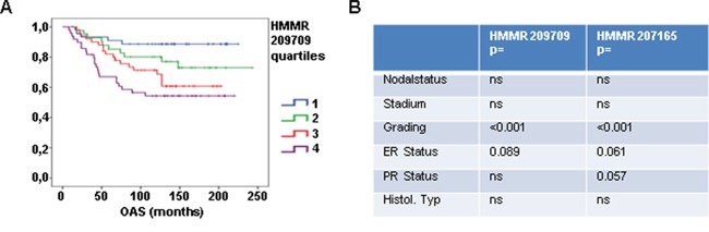
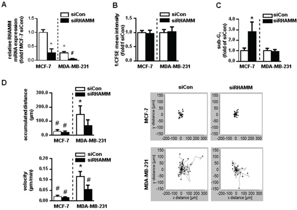
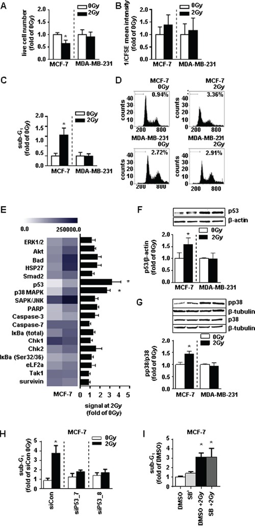
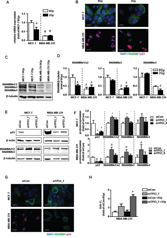
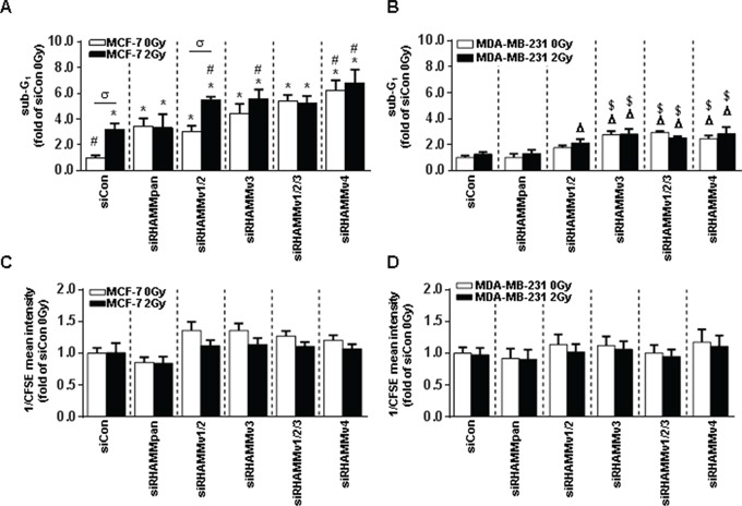
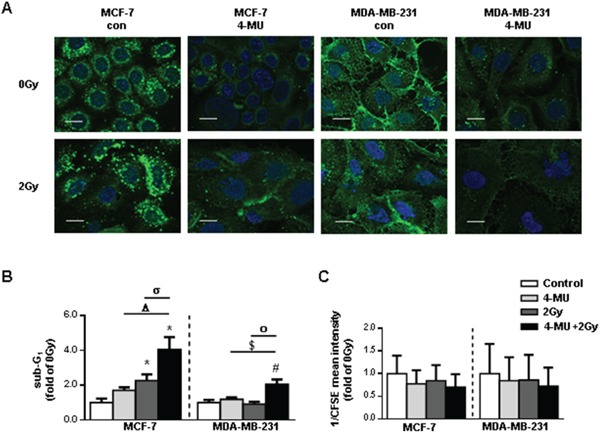
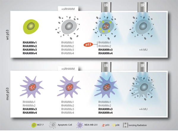
Biomarkers for prognosis in radiotherapy-treated breast cancer patients are urgently needed and important to stratify patients for adjuvant therapies. Recently, a role of the receptor of hyaluronan-mediated motility (RHAMM) has been suggested for tumor progression. Our aim was (i) to investigate the prognostic value of RHAMM in breast cancer and (ii) to unravel its potential function in the radiosusceptibility of breast cancer cells. We demonstrate that RHAMM mRNA expression in breast cancer biopsies is inversely correlated with tumor grade and overall survival. Radiosusceptibility in vitro was evaluated by sub-G1 analysis (apoptosis) and determination of the proliferation rate. The potential role of RHAMM was addressed by short interfering RNAs against RHAMM and its splice variants. High expression of RHAMMv1/v2 in p53 wild type cells (MCF-7) induced cellular apoptosis in response to ionizing radiation. In comparison, in p53 mutated cells (MDA-MB-231) RHAMMv1/v2 was expressed sparsely resulting in resistance towards irradiation induced apoptosis. Proliferation capacity was not altered by ionizing radiation in both cell lines. Importantly, pharmacological inhibition of the major ligand of RHAMM, hyaluronan, sensitized both cell lines towards radiation induced cell death. Based on the present data, we conclude that the detection of RHAMM splice variants in correlation with the p53 mutation status could help to predict the susceptibility of breast cancer cells to radiotherapy. Additionally, our studies raise the possibility that the response to radiotherapy in selected cohorts may be improved by pharmaceutical strategies against RHAMM and its ligand hyaluronan.

Keywords: RHAMM; breast cancer; cell death; extracellular matrix; ionizing radiation.


Conflict of interest statement

The authors declare no conflicts of interest.

Figures

Figure 1
Figure 1. RHAMM is prognostic for patient overall survival
A. Affymetrix analysis of RHAMM expression in 196 tissue samples from breast cancer patients is shown. Patients were stratified into subgroups according their RHAMM expression (low (1), medium (2), high (3), very high (4)) and the subgroups were correlated to overall survival. B. table showing results of statistic tests for clinical parameter in two affymetrix analysis.
Figure 2
Figure 2. RHAMM has apoptotic and motility characteristics in different cancer cell lines in vitro
A. relative mRNA expression of RHAMM in siRHAMM transfected MCF-7 and MDA-MB-231 cells. B. proliferation rate measured with CFSE staining in MCF-7 and MDA-MB-231 cells 48h after siRNA knockdown of RHAMM. CFSE intensity is assigned reciprocally. C. sub-G1 analysis of MCF-7 and MDA-MB-231 cells 48h after siRNA knockdown of RHAMM. D. accumulated distance and velocity of migration assay with exemplary pictures of one experiment. *, p<0.05 in comparison to MCF-7 siCon; #, p<0.05 in comparison to MDA-MB-231 siCon.
Figure 3
Figure 3. MDA-MB-231 cells are not radiosensitive at a dose of 2Gy whereas MCF-7 cells are radiosensitive via activation of p53 and p38
A. live cell number, B. proliferation rate of via CFSE (assigned reciprocally), and C. sub-G1 analysis of MCF-7 and MDA-MB-231 cells 48h after irradiation with 2Gy measurement. D. representative histograms of sub-G1 analyses. E. absolute expression levels of proteins analyzed via the stress and apoptosis assay depicted in a heatmap (left) and the fold changes of these proteins in MCF-7 cells 48h after irradiation compared to the non-irradiated control (right). F. western blot analyses of MCF-7 cell lysates for p53 and G. pp38 and p38 protein expression. H. sub-G1 analysis in MCF-7 cells transfected with siRNA against p53 48h after irradiation with 2Gy. I. proliferation rate of MCF-7 cells treated with SB (SB202190, p38-inhibitor, 10 μM) 4h before irradiation with 2Gy. *, p<0.05 in comparison with untreated MCF-7 0Gy (A-G, J, K)/siCon (H, I).
Figure 4
Figure 4. p53 and RHAMM variant status of MCF-7 and MDA-MB-231 in response to 2Gy irradiation
A. relative mRNA expression of RHAMMpan. B. RHAMM (green) and p53 (pink) immunofluorescence staining 48h after irradiation with 2Gy. Scale bars: 20μm. C. western blot analysis of RHAMM protein expression and its quantification and D. quantification. E. exemplary blots of protein expression of p53 and RHAMM of p53 depleted MCF-7 and MDA-MB-231 cells and F. quantification. G. RHAMM (green) and p53 (pink) immunofluorescence staining 48h after siRNA knockdown of p53. Scale bars: 20μm. H. MDA-MB-231 were transfected with sip53. 48h after transfection cells were irradiated. Sub-G1 was analyzed further 48h later.*, p<0.05 in comparison to MCF-7 siCon, #, p<0.05 MDA-MB-231 in comparison to MCF-7 siCon.
Figure 5
Figure 5. RHAMM pan and RHAMM variant knock-down in MCF-7 and MDA-MB-231 cells leads to increased radiosensitivity after irradiation with 2Gy
A. sub-G1 cell cycle analysis of siRHAMM variants transfected MCF-7 and B. MDA-MB-231 cells 48h after irradiation with 2Gy. C. analysis of proliferation rate via CFSE measurement (assigned reciprocally) in siRHAMM variant transfected MCF-7 and D. MDA-MB-231 cells 48h after irradiation with 2Gy. *, p<0.05 in comparison to MCF-7 siCon 0Gy, #, p<0.05 in comparison to MCF-7 siCon 2Gy, σ, p<0.05 MCF-7 siRHAMMv1/2 2Gy in comparison to MCF-7 siRHAMMv1/2 0Gy, Δ, p<0.05 in comparison to MDA-MB-231 siCon 0Gy, $, p<0.05 in comparison to MDA-MB-231 siCon 2Gy.
Figure 6
Figure 6. Pharmacological inhibition of HA system via 4-MU in MCF-7 and MDA-MB-231 leads to increased radiosensitivity
A. affinitycytochemistry of HA 48h after irradiation with 2Gy. Scale bars: 20μm. B. sub-G1 cell cycle analysis of 4-MU treated MCF-7 and MDA-MB-231 cells 48h after irradiation with 2Gy. C. analysis of proliferation rate via CFSE staining of 4-MU treated MCF-7 and MDA-MB-231 cells 48h after irradiation with 2Gy. CFSE intensity is assigned reciprocally. *, p<0.05 in comparison to MCF-7 0Gy, Δ, p<0.05 in comparison to MCF-7 4-MU, σ, p<0.05 in comparison to MCF-7 2Gy, #, p<0.05 in comparison to MCF-7 0Gy, $, p<0.05 in comparison to MCF-7 4-MU, ο, p<0.05 in comparison to MCF-7 2Gy.
Figure 7
Figure 7. Schematic model of mamma-ca radiosensitization
Breast cancer cells with wildtype p53 status are radiosensitive and can be forced into apoptosis upon RHAMM downregulation. Mutant p53 breast cancer cells are only radiosensitive if treated with 4-MU which affects RHAMM ligand inhibition.

Similar articles

Cited by

References

    1. Favourable and unfavourable effects on long-term survival of radiotherapy for early breast cancer: an overview of the randomised trials. Lancet. 2000;355:1757–1770. - PubMed
    1. Mokbel K. Current management of ductal carcinoma in situ of the breast. Int. J. Clin. Oncol. 2003;8:18–22. - PubMed
    1. Boyages J, Delaney G, Taylor R. Predictors of local recurrence after treatment of ductal carcinoma in situ: a meta-analysis. Cancer. 1999;85:616–28. - PubMed
    1. Hooning MJ, Botma A, Aleman BM, Baaijens MH, Bartelink H, Klijn JG, Taylor CW, van Leeuwen FE. Long-term risk of cardiovascular disease in 10-year survivors of breast cancer. J. Natl. Cancer Inst. 2007;99:365–75. - PubMed
    1. Wilder RB, Curcio LD, Khanijou RK, Eisner ME, Kakkis JL, Chittenden L, Agustin J, Lizarde J, Mesa AV, Macedo JC, Ravera J, Tokita KM. Results with accelerated partial breast irradiation in terms of estrogen receptor, progesterone receptor, and human growth factor receptor 2 status. Int. J. Radiat. Oncol. Biol. Phys. 2010;78:799–803. - PubMed

MeSH terms

Substances