

The status of epidermal growth factor receptor in borderline ovarian tumours
- PMID: 26870997
- PMCID: PMC4891141
- DOI: 10.18632/oncotarget.7257
The status of epidermal growth factor receptor in borderline ovarian tumours
Abstract
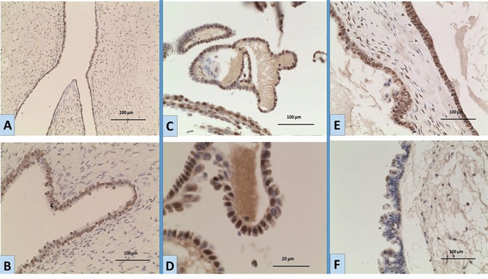
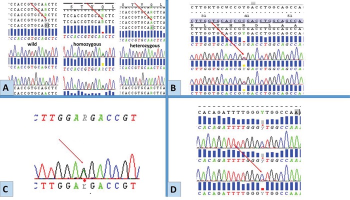
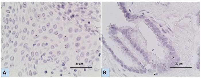
The majority of borderline ovarian tumours (BOTs) behave in a benign fashion, but some may show aggressive behavior. The reason behind this has not been elucidated. The epidermal growth factor receptor (EGFR) is known to contribute to cell survival signals as well as metastatic potential of some tumours. EGFR expression and gene status have not been thoroughly investigated in BOTs as it has in ovarian carcinomas. In this study we explore protein expression as well as gene mutations and amplifications of EGFR in BOTs in comparison to a subset of other epithelial ovarian tumours. We studied 85 tumours, including 61 BOTs, 10 low grade serous carcinomas (LGSCs), 9 high grade serous carcinomas (HGSCs) and 5 benign epithelial tumours. EGFR protein expression was studied using immunohistochemistry. Mutations were investigated by Sanger sequencing exons 18-21 of the tyrosine kinase domain of EGFR. Cases with comparatively higher protein expression were examined for gene amplification by chromogenic in situ hybridization. We also studied the tumours for KRAS and BRAF mutations. Immunohistochemistry results revealed both cytoplasmic and nuclear EGFR expression with variable degrees between tumours. The level of nuclear localization was relatively higher in BOTs and LGSCs as compared to HGSCs or benign tumours. The degree of nuclear expression of BOTs showed no significant difference from that in LGSCs (mean ranks 36.48, 33.05, respectively, p=0.625), but was significantly higher than in HGSCs (mean ranks: 38.88, 12.61 respectively, p< 0.001) and benign tumours (mean ranks: 35.18, 13.00 respectively, p= 0.010). Cytoplasmic expression level was higher in LGSCs. No EGFR gene mutations or amplification were identified, yet different polymorphisms were detected. Five different types of point mutations in the KRAS gene and the V600E BRAF mutation were detected exclusively in BOTs and LGSCs. Our study reports for the first time nuclear localization of EGFR in BOTs. The nuclear localization similarities between BOTs and LGSCs and not HGSCs support the hypothesis suggesting evolution of LGSCs from BOTs. We also confirm that EGFR mutations and amplifications are not molecular events in the pathogenesis of BOTs.
Keywords: EGFR; borderline ovarian tumours.
Conflict of interest statement
The authors declare no conflicts of interest
Figures
Similar articles
-
KRAS (but not BRAF) mutations in ovarian serous borderline tumour are associated with recurrent low-grade serous carcinoma.J Pathol. 2013 Dec;231(4):449-56. doi: 10.1002/path.4252. J Pathol. 2013. PMID: 24549645 Free PMC article.
-
BRAFV600E mutations and immunohistochemical expression of VE1 protein in low-grade serous neoplasms of the ovary.Histopathology. 2018 Sep;73(3):438-443. doi: 10.1111/his.13651. Epub 2018 Jun 22. Histopathology. 2018. PMID: 29770477 Free PMC article.
-
Mutational analysis of BRAF and KRAS in ovarian serous borderline (atypical proliferative) tumours and associated peritoneal implants.J Pathol. 2014 Jan;232(1):16-22. doi: 10.1002/path.4293. J Pathol. 2014. PMID: 24307542 Free PMC article.
-
Biomolecular pathogenesis of borderline ovarian tumors: focusing target discovery through proteogenomics.Curr Cancer Drug Targets. 2010 Feb;10(1):107-16. doi: 10.2174/156800910790980269. Curr Cancer Drug Targets. 2010. PMID: 20088785 Review.
-
Low Grade Serous Ovarian Carcinoma: from the molecular characterization to the best therapeutic strategy.Cancer Treat Rev. 2015 Feb;41(2):136-43. doi: 10.1016/j.ctrv.2014.12.003. Epub 2014 Dec 23. Cancer Treat Rev. 2015. PMID: 25573350 Review.
Cited by
-
Two missense variants of the epidermal growth factor receptor gene are associated with non small cell lung carcinoma in the subjects from Iraq.Mol Biol Rep. 2022 Dec;49(12):11653-11661. doi: 10.1007/s11033-022-07933-w. Epub 2022 Sep 28. Mol Biol Rep. 2022. PMID: 36169894
-
Rethink of EGFR in Cancer With Its Kinase Independent Function on Board.Front Oncol. 2019 Aug 23;9:800. doi: 10.3389/fonc.2019.00800. eCollection 2019. Front Oncol. 2019. PMID: 31508364 Free PMC article.
-
Loss of 1p36.33 Frequent in Low-Grade Serous Ovarian Cancer.Neoplasia. 2019 Jun;21(6):582-590. doi: 10.1016/j.neo.2019.03.014. Epub 2019 May 1. Neoplasia. 2019. PMID: 31054497 Free PMC article.
-
Nuclear epidermal growth factor receptor (nEGFR) in clinical treatment.Heliyon. 2024 Nov 5;10(21):e40150. doi: 10.1016/j.heliyon.2024.e40150. eCollection 2024 Nov 15. Heliyon. 2024. PMID: 39568844 Free PMC article. Review.
-
Evaluation of vitamin D biosynthesis and pathway target genes reveals UGT2A1/2 and EGFR polymorphisms associated with epithelial ovarian cancer in African American Women.Cancer Med. 2019 May;8(5):2503-2513. doi: 10.1002/cam4.1996. Epub 2019 Apr 18. Cancer Med. 2019. PMID: 31001917 Free PMC article.
References
-
- Lee KR. Tumours of the ovary and peritoneum. In: Tavassoli FA, Devilee P, World Health Organization classification of tumours, editors. Pathology and genetics of tumours of the breast and female genital organs. Lyon: IARC Press; 2003. pp. 114–133.
-
- Daraï E, Fauvet R, Uzan C, Gouy S, Duvillard P, Morice P. Fertility and borderline ovarian tumor: a systematic review of conservative management, risk of recurrence and alternative options. Hum Reprod Update. 2013;19:151–166. - PubMed
-
- Shigematsu H, Lin L, Takahashi T, Nomura M, Suzuki M, Wistuba II, Fong KM, Lee H, Toyooka S, Shimizu N, Fujisawa T, Feng Z, Roth JA, et al. Clinical and biological features associated with epidermal growth factor receptor gene mutations in lung cancers. J Natl Cancer Inst. 2005;97:339–346. - PubMed
-
- Shih IeM, Kurman RJ. Molecular pathogenesis of ovarian borderline tumors: new insights and old challenges. Clin Cancer Res. 2005;11:7273–7279. - PubMed
-
- Yewale C, Baradia D, Vhora I, Patil S, Misra A. Epidermal growth factor receptor targeting in cancer: a review of trends and strategies. biomaterials. 2013;34:8690–8707. - PubMed
Publication types
MeSH terms
Substances
Grants and funding
LinkOut - more resources
Full Text Sources
Other Literature Sources
Medical
Research Materials
Miscellaneous
