. 2016 May;20(5):949-61.
doi: 10.1111/jcmm.12795. Epub 2016 Feb 12.

Simvastatin improves the homing of BMSCs via the PI3K/AKT/miR-9 pathway

Affiliations

Simvastatin improves the homing of BMSCs via the PI3K/AKT/miR-9 pathway

Weidong Bing et al. J Cell Mol Med. 2016 May.

Abstract

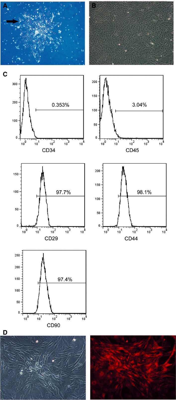
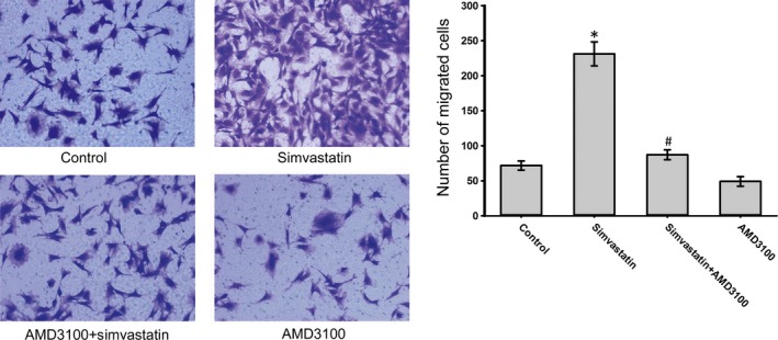
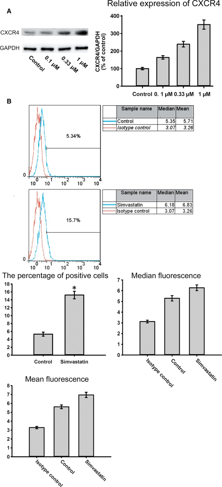
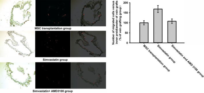
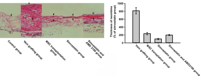
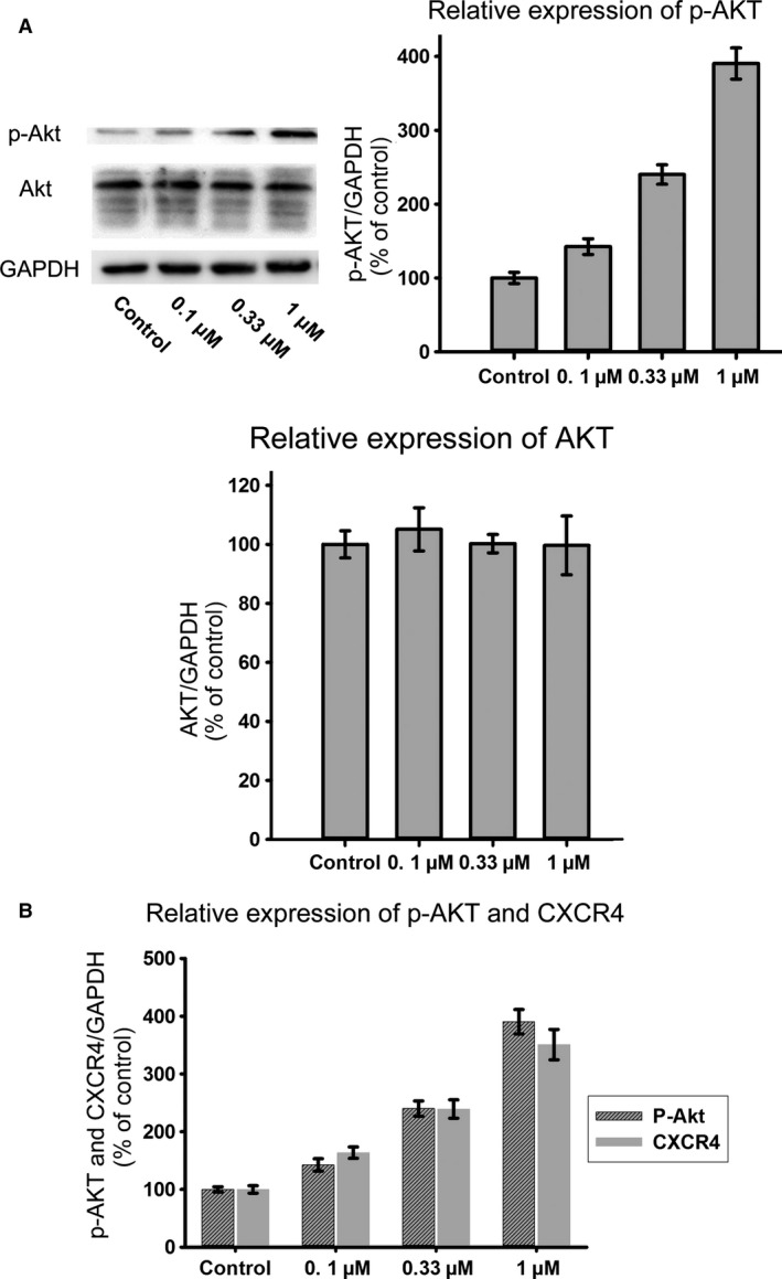
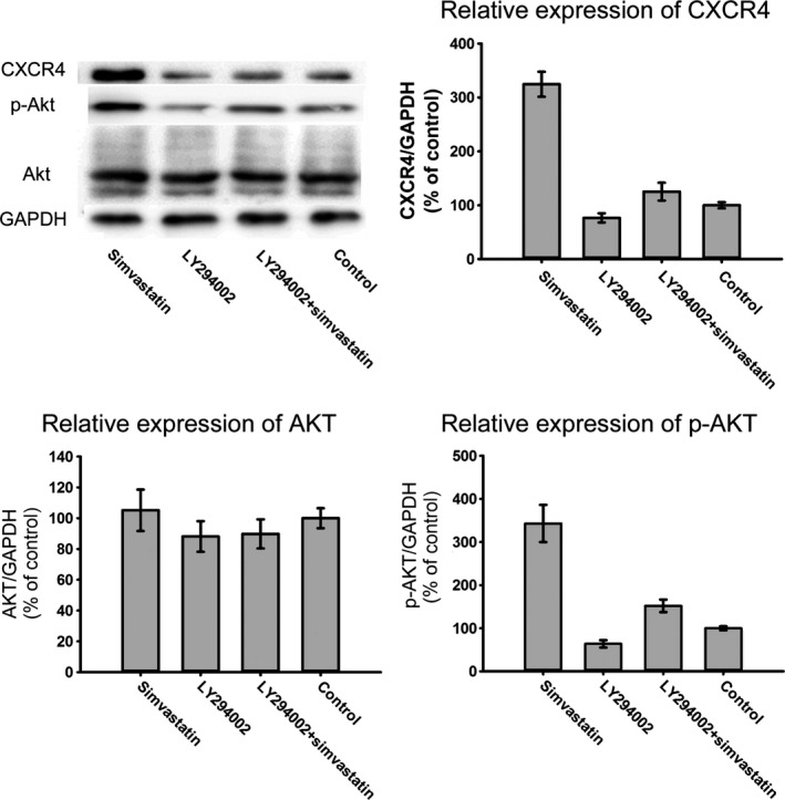
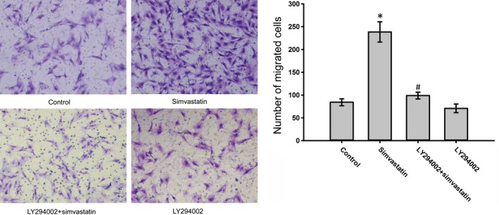
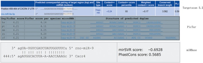
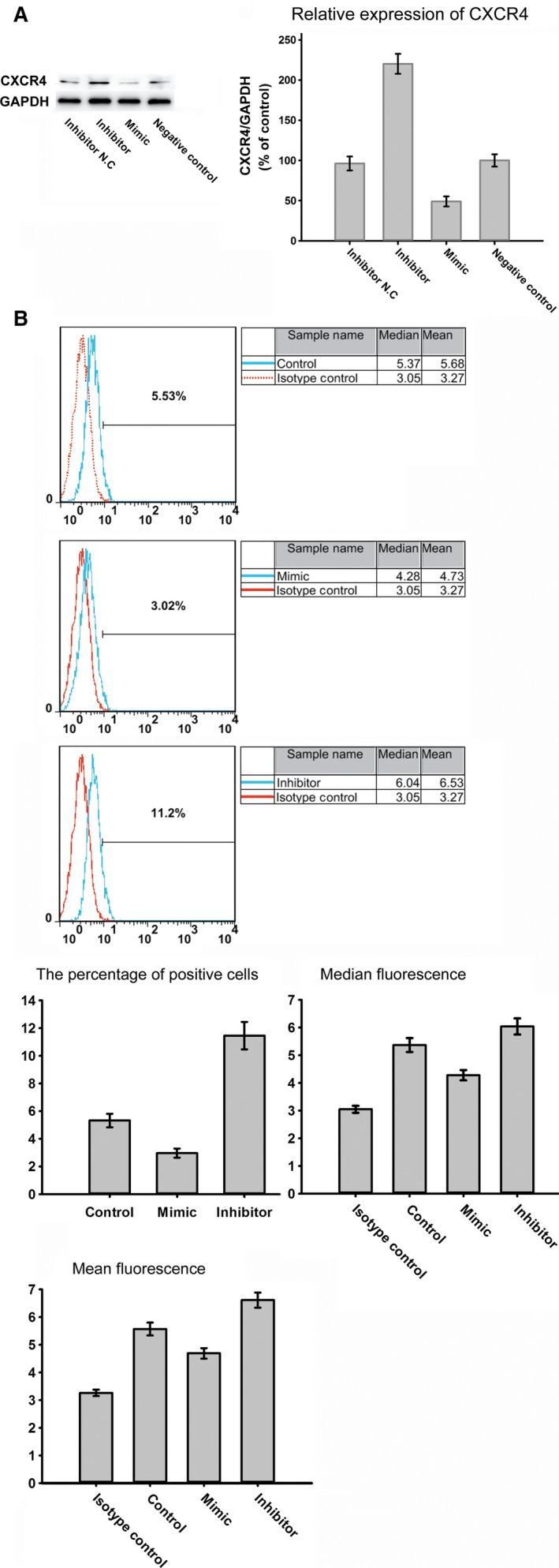
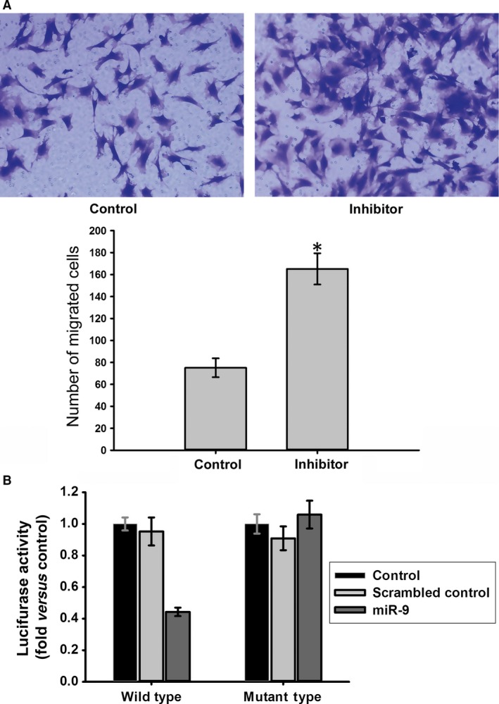
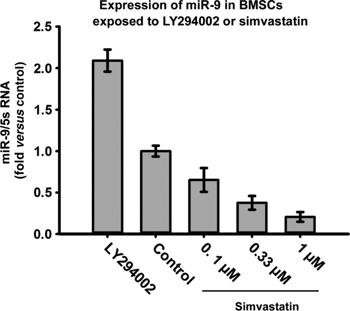
Bone marrow-derived mesenchymal stem cells (BMSCs) have great therapeutic potential for many diseases. However, the homing of BMSCs to injury sites remains a difficult problem. Recent evidence indicates that simvastatin stimulates AKT phosphorylation, and p-AKT affects the expression of chemokine (CXC motif) receptor-4 (CXCR4). Therefore, simvastatin may improve the expression of CXCR4 in BMSCs, and microRNAs (miRs) may participate in this process. In this study, we demonstrated that simvastatin increased both the total and the surface expression of CXCR4 in BMSCs. Stromal cell-derived factor-1α (SDF-1α)-induced migration of BMSCs was also enhanced by simvastatin, and this action was inhibited by AMD 3100(a chemokine receptor antagonist for CXCR4). The PI3K/AKT pathway was activated by simvastatin in this process, and LY294002 reversed the overexpression of CXCR4 caused by simvastatin. MiR-9 directly targeted CXCR4 in rat BMSCs, and simvastatin decreased miR-9 expression. P-AKT affected the expression of miR-9; as the phosphorylation of AKT increased, miR-9 expression decreased. In addition, LY294002 increased miR-9 expression. Taken together, our results indicated that simvastatin improved the migration of BMSCs via the PI3K/AKT pathway. MiR-9 also participated in this process, and the phosphorylation of AKT affected miR-9 expression, suggesting that simvastatin might have beneficial effects in stem cell therapy.

Keywords: AKT; BMSCs; CXCR4; microRNA; simvastatin.


Figures

Figure 1
Figure 1
Characterization of BMSCs. (A) BMSCs exhibited a fibroblast–like shape and forms a cell colony (72 h after isolation, ×40. And the arrow indicates the cell colony). (B) Undifferentiated BMSCs after three passages of culture, display a flattened fibroblast‐like morphology (C). Flow cytometric analysis of cultured MSCs with CD29, CD44, CD90, CD34 and CD45 antibodies. BMSCs used in our study express CD90 and CD44, showing their mesenchymal origin. (D) Labelled cells showed red fluorescence under excitation.
Figure 2
Figure 2
Simvastatin improves SDF‐1α‐induced migration of BMSCs. (A) Untreated BMSCs (Control), BMSCs treated with simvastatin (Simvastatin), and BMSCs pre‐treated with AMD3100 prior to simvastatin (AMD3100 + simvastatin) were placed in the upper chamber. The migrated cells were observed under a microscope (×200) *P < 0.05, versus Control group; # P < 0.05, compared with the Simvastatin group.
Figure 3
Figure 3
(A) The expression level of CXCR4 in BMSCs under the effect of different concentrations of simvastatin. The ratio of the expression of CXCR4 in different groups to that of the control group was calculated. (B) The surface expression of CXCR4 in the BMSCs increased following treatment with simvastatin.
Figure 4
Figure 4
Morphology of frozen sections of each group was showed in the left picture. The fluorescence of labelled cells was showed in the middle one and the right one was formed by merging the first two pictures. Labelled BMSCs were more in simvastatin group in vein grafts with similar inner perimeter than MSC transplantation group and simvastatin and AMD 3100 group.
Figure 5
Figure 5
HE staining sections of vein grafts from week 4 after operation were analysed for neointima formation. The arrows indicate the control vessel wall in control group and neointima in the rest groups. Formation of neointima of vein grafts was most reduced in simvastatin group.
Figure 6
Figure 6
(A) Different concentrations of simvastatin led to different expression levels of p‐AKT in BMSCs; however expression of AKT showed no significant change. (B) As the concentration of simvastatin increased, expression of p‐AKT and CXCR4 increased synchronously.
Figure 7
Figure 7
Western blot analysis of the protein expression levels of CXCR4, p‑AKT and AKT in BMSCs treated with simvastatin and/or LY294002. Treatment of LY294002 could partially inhibit phosphorylation of AKT and expression of CXCR4.
Figure 8
Figure 8
Effects of simvastatin and/or LY294002 on the migration of BMSCs to SDF‑1α. LY294002 could reduce the improvement of migration caused by simvastatin. The migrated cells were observed under a microscope (×200). *P < 0.05, versus Control group; # P < 0.05, compared with the Simvastatin group.
Figure 9
Figure 9
MiR‐9 might target CXCR4 with good scores according to TargetScan 5.2, the online miRNA prediction tool of the PicTar, and miRBase.
Figure 10
Figure 10
(A) The expression level of CXCR4 in BMSCs after transfection with inhibitor, Inhibitor N. C, mimic and negative control. (B) The surface expression of CXCR4 in the BMSCs following transfection.
Figure 11
Figure 11
(A) Effects of miR‐9 on the migration of BMSCs to SDF‑1α. Transfection with inhibitor led to an up‐regulation of migration. *P < 0.05, versus Control group (B) Luciferase activity was suppressed by miR‐9, but not scrambled control in the wild‐type reporter group. In the mutant‐type reporter group, luciferase activity showed no significant difference under different treatment. The wild‐type reporter contains the full‐length 3′UTR of rat CXCR4 mRNA including the sequence complementary to miR‐9 and this sequence is mutated in mutant‐type reporter. *P < 0.05, versus Control group.
Figure 12
Figure 12
Expression of miR‐9 might be related to the phosphorylation of AKT. As the concentration of simvastatin increased, expression of mir‐9 decreased which was opposite to the trend of p‐AKT. LY294002 could increase expression of mir‐9.

References

    1. Fisher SA, Brunskill SJ, Doree C, et al Stem cell therapy for chronic ischaemic heart disease and congestive heart failure. Cochrane Database Syst Rev. 2014; 4: Cd007888. - PubMed
    1. Ko IK, Kim BS. Mesenchymal stem cells for treatment of myocardial infarction. Int J Stem Cells. 2008; 1: 49–54. - PMC - PubMed
    1. Yang Z, Zhu L, Li F, et al Bone marrow stromal cells as a therapeutic treatment for ischemic stroke. Neurosci Bull. 2014; 30: 524–34. - PMC - PubMed
    1. Stroncek DF, Sabatino M, Ren J, et al Establishing a bone marrow stromal cell transplant program at the National Institutes of Health Clinical Center. Tissue Eng Part B Rev. 2014; 20: 200–5. - PMC - PubMed
    1. Vaquero J, Zurita M. Bone marrow stromal cells for spinal cord repair: a challenge for contemporary neurobiology. Histol Histopathol. 2009; 24: 107–16. - PubMed

Publication types

MeSH terms

LinkOut - more resources