

Estrogen receptor-mediated miR-486-5p regulation of OLFM4 expression in ovarian cancer
- PMID: 26871282
- PMCID: PMC4891143
- DOI: 10.18632/oncotarget.7236
Estrogen receptor-mediated miR-486-5p regulation of OLFM4 expression in ovarian cancer
Abstract
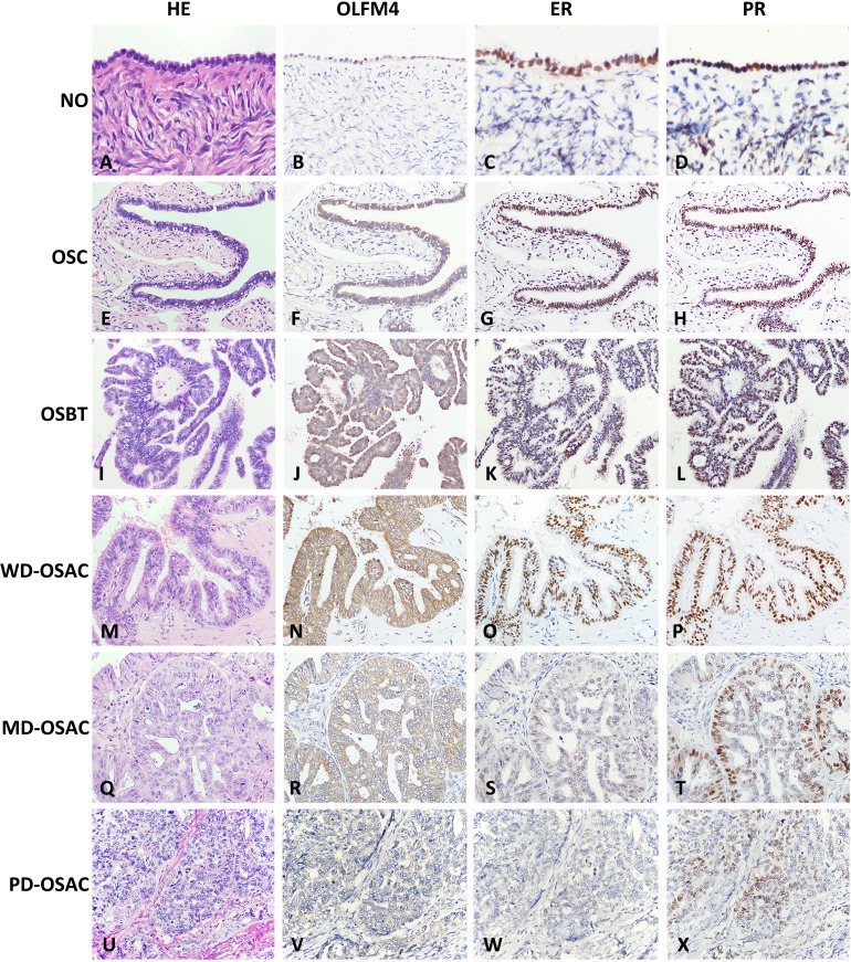
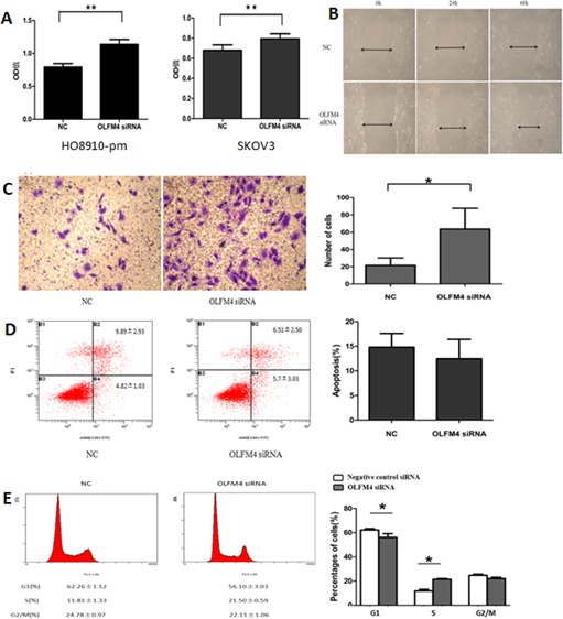
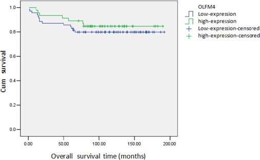
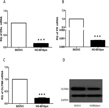
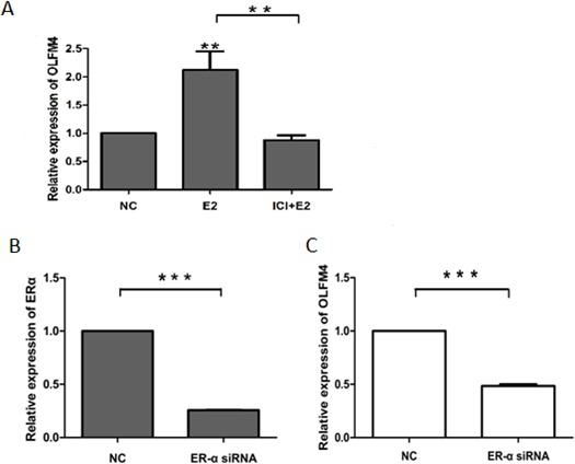
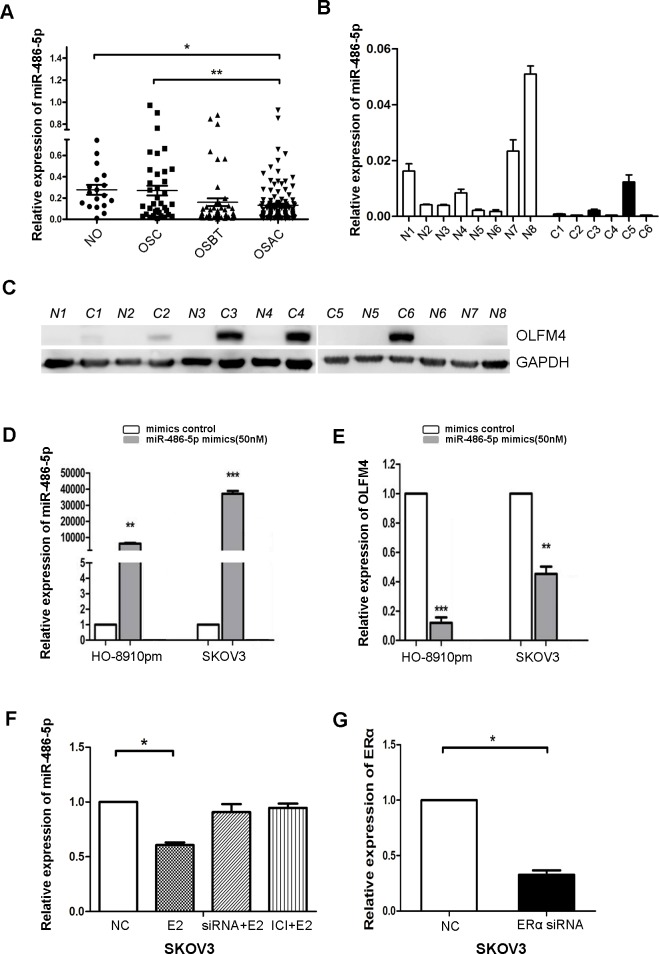
Estrogen signaling influences the development and progression of ovarian tumors, but the underlying mechanisms are not well understood. In a previous study we demonstrated that impairment of estrogen receptor alpha (ERα)-mediated olfactomedin 4 (OLFM4) expression promotes the malignant progression of endometrioid adenocarcinoma, and we identified OLFM4 as a potential target of miR-486-5p. In this study we investigated the role of OLFM4 in ovarian serous adenocarcinoma. Ovarian serous adenocarcinoma tissues had reduced OLFM4 expression. Expression of OLFM4 was positively correlated with ERα expression, and estrogen (E2) treatment in ovarian cancer cells induced OLFM4 expression in an ERα-dependent manner. In contrast to ERα, miR-486-5p levels were inversely correlated with OLFM4 expression in ovarian serous adenocarcinoma. Ovarian cancer cells transfected with miR-486-5p mimics showed decreased OLFM4 mRNA expression, and ovarian cancer cells treated with E2 showed reduced cellular miR-486-5p levels. OLFM4 knockdown enhanced proliferation, migration, and invasion by ovarian cancer cells. Low expression of OLFM4 was also associated with high tumor FIGO stage and poor tumor differentiation. These results suggest OLFM4 is downregulated by miR-486-5p, which contributes to ovarian cancer tumorigenesis. Conversely, estrogen receptor signaling downregulates miR-486-5p and upregulates OLFM4 expression, slowing the development and progression of ovarian cancer.
Keywords: OLFM4; estrogen; miR-486-5p; ovarian serous adenocarcinoma.
Conflict of interest statement
The authors confirm that there are no conflicts of interest.
Figures
Similar articles
-
Oestrogen receptor-mediated expression of Olfactomedin 4 regulates the progression of endometrial adenocarcinoma.J Cell Mol Med. 2014 May;18(5):863-74. doi: 10.1111/jcmm.12232. Epub 2014 Feb 4. J Cell Mol Med. 2014. PMID: 24495253 Free PMC article.
-
RhoC is a major target of microRNA-93-5P in epithelial ovarian carcinoma tumorigenesis and progression.Mol Cancer. 2015 Feb 4;14(1):31. doi: 10.1186/s12943-015-0304-6. Mol Cancer. 2015. PMID: 25649143 Free PMC article.
-
MicroRNA-219-5p inhibits the proliferation, migration, and invasion of epithelial ovarian cancer cells by targeting the Twist/Wnt/β-catenin signaling pathway.Gene. 2017 Dec 30;637:25-32. doi: 10.1016/j.gene.2017.09.012. Epub 2017 Sep 7. Gene. 2017. PMID: 28890378
-
Olfactomedin 4 expression and functions in innate immunity, inflammation, and cancer.Cancer Metastasis Rev. 2016 Jun;35(2):201-12. doi: 10.1007/s10555-016-9624-2. Cancer Metastasis Rev. 2016. PMID: 27178440 Review.
-
MiRNA-210: A Current Overview.Anticancer Res. 2017 Dec;37(12):6511-6521. doi: 10.21873/anticanres.12107. Anticancer Res. 2017. PMID: 29187425 Review.
Cited by
-
The Central Roles of Noncoding RNA in Estrogen-Dependent Female Reproductive System Tumors.Int J Endocrinol. 2021 May 24;2021:5572063. doi: 10.1155/2021/5572063. eCollection 2021. Int J Endocrinol. 2021. PMID: 34122542 Free PMC article. Review.
-
High expression of olfactomedin-4 is correlated with chemoresistance and poor prognosis in pancreatic cancer.PLoS One. 2020 Jan 10;15(1):e0226707. doi: 10.1371/journal.pone.0226707. eCollection 2020. PLoS One. 2020. PMID: 31923206 Free PMC article.
-
Olaparib Combined with DDR Inhibitors Effectively Prevents EMT and Affects miRNA Regulation in TP53-Mutated Epithelial Ovarian Cancer Cell Lines.Int J Mol Sci. 2025 Jan 15;26(2):693. doi: 10.3390/ijms26020693. Int J Mol Sci. 2025. PMID: 39859407 Free PMC article.
-
The differential expression of miRNAs between ovarian endometrioma and endometriosis-associated ovarian cancer.J Ovarian Res. 2020 May 2;13(1):51. doi: 10.1186/s13048-020-00652-5. J Ovarian Res. 2020. PMID: 32359364 Free PMC article.
-
Development and validation of prognostic gene signature for basal-like breast cancer and high-grade serous ovarian cancer.Breast Cancer Res Treat. 2020 Dec;184(3):689-698. doi: 10.1007/s10549-020-05884-z. Epub 2020 Sep 2. Breast Cancer Res Treat. 2020. PMID: 32880016 Free PMC article.
References
-
- Cheng EJ, Kurman RJ, Wang M, Oldt R, Wang BG, Berman DM, Shih Ie M. Molecular genetic analysis of ovarian serous cystadenomas. Lab Invest. 2004;84:778–784. - PubMed
-
- O'Donnell AJ, Macleod KG, Burns DJ, Smyth JF, Langdon SP. Estrogen receptor-alpha mediates gene expression changes and growth response in ovarian cancer cells exposed to estrogen. Endocr Relat Cancer. 2005;12:851–866. - PubMed
-
- Lindgren P, Backstrom T, Mahlck CG, Ridderheim M, Cajander S. Steroid receptors and hormones in relation to cell proliferation and apoptosis in poorly differentiated epithelial ovarian tumors. Int J Oncol. 2001;19:31–38. - PubMed
-
- Burges A, Bruning A, Dannenmann C, Blankenstein T, Jeschke U, Shabani N, Friese K, Mylonas I. Prognostic significance of estrogen receptor alpha and beta expression in human serous carcinomas of the ovary. Arch Gynecol Obstet. 2010;281:511–517. - PubMed
Publication types
MeSH terms
Substances
LinkOut - more resources
Full Text Sources
Other Literature Sources
Medical
Miscellaneous
