

Lipid tethering of breast tumor cells enables real-time imaging of free-floating cell dynamics and drug response
- PMID: 26871289
- PMCID: PMC4891134
- DOI: 10.18632/oncotarget.7251
Lipid tethering of breast tumor cells enables real-time imaging of free-floating cell dynamics and drug response
Abstract
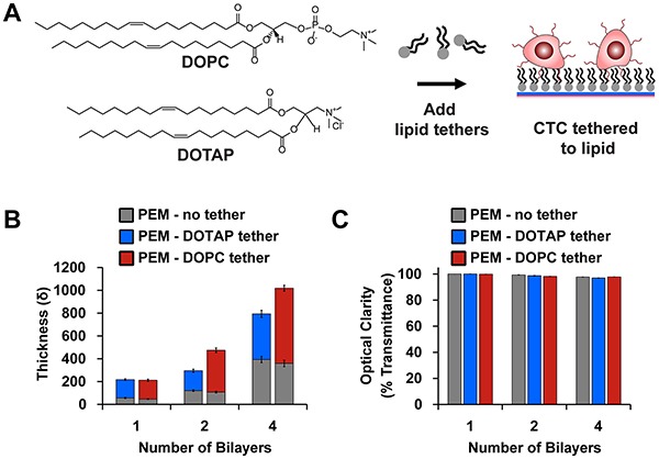
Free-floating tumor cells located in the blood of cancer patients, known as circulating tumor cells (CTCs), have become key targets for studying metastasis. However, effective strategies to study the free-floating behavior of tumor cells in vitro have been a major barrier limiting the understanding of the functional properties of CTCs. Upon extracellular-matrix (ECM) detachment, breast tumor cells form tubulin-based protrusions known as microtentacles (McTNs) that play a role in the aggregation and re-attachment of tumor cells to increase their metastatic efficiency. In this study, we have designed a strategy to spatially immobilize ECM-detached tumor cells while maintaining their free-floating character. We use polyelectrolyte multilayers deposited on microfluidic substrates to prevent tumor cell adhesion and the addition of lipid moieties to tether tumor cells to these surfaces through interactions with the cell membranes. This coating remains optically clear, allowing capture of high-resolution images and videos of McTNs on viable free-floating cells. In addition, we show that tethering allows for the real-time analysis of McTN dynamics on individual tumor cells and in response to tubulin-targeting drugs. The ability to image detached tumor cells can vastly enhance our understanding of CTCs under conditions that better recapitulate the microenvironments they encounter during metastasis.
Keywords: breast cancer; circulating tumor cells; microfluidics; microtentacles; polyelectrolyte multilayers.
Conflict of interest statement
The University of Maryland has filed patents on which J. I. Andorko, K. R. Chakrabarti, C. M. Jewell, S.S. Martin, E. L. Sooklal, and R.A. Whipple are listed as inventors. The other authors have no conflicts of interests to disclose.
Figures


Similar articles
-
Partial thermal imidization of polyelectrolyte multilayer cell tethering surfaces (TetherChip) enables efficient cell capture and microtentacle fixation for circulating tumor cell analysis.Lab Chip. 2020 Aug 21;20(16):2872-2888. doi: 10.1039/d0lc00207k. Epub 2020 Aug 3. Lab Chip. 2020. PMID: 32744284 Free PMC article.
-
ROCK inhibition promotes microtentacles that enhance reattachment of breast cancer cells.Oncotarget. 2015 Mar 20;6(8):6251-66. doi: 10.18632/oncotarget.3360. Oncotarget. 2015. PMID: 25749040 Free PMC article.
-
Curcumin targets breast cancer stem-like cells with microtentacles that persist in mammospheres and promote reattachment.Cancer Res. 2014 Feb 15;74(4):1250-60. doi: 10.1158/0008-5472.CAN-13-1778. Epub 2013 Dec 26. Cancer Res. 2014. PMID: 24371229 Free PMC article.
-
Microtentacles tip the balance of cytoskeletal forces in circulating tumor cells.Cancer Res. 2010 Oct 15;70(20):7737-41. doi: 10.1158/0008-5472.CAN-10-1569. Epub 2010 Oct 5. Cancer Res. 2010. PMID: 20924109 Free PMC article. Review.
-
Breast cancer metastasis.Cancer Genomics Proteomics. 2012 Sep-Oct;9(5):311-20. Cancer Genomics Proteomics. 2012. PMID: 22990110 Review.
Cited by
-
Engineering Cell Surfaces with Polyelectrolyte Materials for Translational Applications.Polymers (Basel). 2017 Jan 28;9(2):40. doi: 10.3390/polym9020040. Polymers (Basel). 2017. PMID: 30970718 Free PMC article. Review.
-
Tubulin-Based Microtentacles Aid in Heterotypic Clustering of Neutrophil-Differentiated HL-60 Cells and Breast Tumor Cells.Adv Sci (Weinh). 2025 Feb;12(6):e2409260. doi: 10.1002/advs.202409260. Epub 2024 Dec 18. Adv Sci (Weinh). 2025. PMID: 39696759 Free PMC article.
-
Partial thermal imidization of polyelectrolyte multilayer cell tethering surfaces (TetherChip) enables efficient cell capture and microtentacle fixation for circulating tumor cell analysis.Lab Chip. 2020 Aug 21;20(16):2872-2888. doi: 10.1039/d0lc00207k. Epub 2020 Aug 3. Lab Chip. 2020. PMID: 32744284 Free PMC article.
-
Insights on CTC Biology and Clinical Impact Emerging from Advances in Capture Technology.Cells. 2019 Jun 6;8(6):553. doi: 10.3390/cells8060553. Cells. 2019. PMID: 31174404 Free PMC article. Review.
-
Extracting microtentacle dynamics of tumor cells in a non-adherent environment.Oncotarget. 2017 Dec 4;8(67):111567-111580. doi: 10.18632/oncotarget.22874. eCollection 2017 Dec 19. Oncotarget. 2017. PMID: 29340075 Free PMC article.
References
-
- Cristofanilli M, Budd GT, Ellis MJ, Stopeck A, Matera J, Miller MC, Reuben JM, Doyle GV, Allard WJ, Terstappen LW, Hayes DF. Circulating tumor cells, disease progression, and survival in metastatic breast cancer. N Engl J Med. 2004;351:781–791. - PubMed
-
- Krebs MG, Sloane R, Priest L, Lancashire L, Hou JM, Greystoke A, Ward TH, Ferraldeschi R, Hughes A, Clack G, Ranson M, Dive C, Blackhall FH. Evaluation and prognostic significance of circulating tumor cells in patients with non-small-cell lung cancer. J Clin Oncol. 2011;29:1556–1563. - PubMed
-
- Stott SL, Hsu CH, Tsukrov DI, Yu M, Miyamoto DT, Waltman BA, Rothenberg SM, Shah AM, Smas ME, Korir GK, Floyd FP, Jr, Gilman AJ, Lord JB, Winokur D, Springer S, Irimia D, et al. Isolation of circulating tumor cells using a microvortex-generating herringbone-chip. Proc Natl Acad Sci U S A. 2010;107:18392–18397. - PMC - PubMed
Publication types
MeSH terms
Substances
Grants and funding
LinkOut - more resources
Full Text Sources
Other Literature Sources
Medical
Molecular Biology Databases
