. 2016 Feb 12;11(2):e0149064.
doi: 10.1371/journal.pone.0149064. eCollection 2016.

Hepatitis C Virus Resistance to Carbohydrate-Binding Agents

Affiliations

Hepatitis C Virus Resistance to Carbohydrate-Binding Agents

Laure Izquierdo et al. PLoS One. .

Abstract

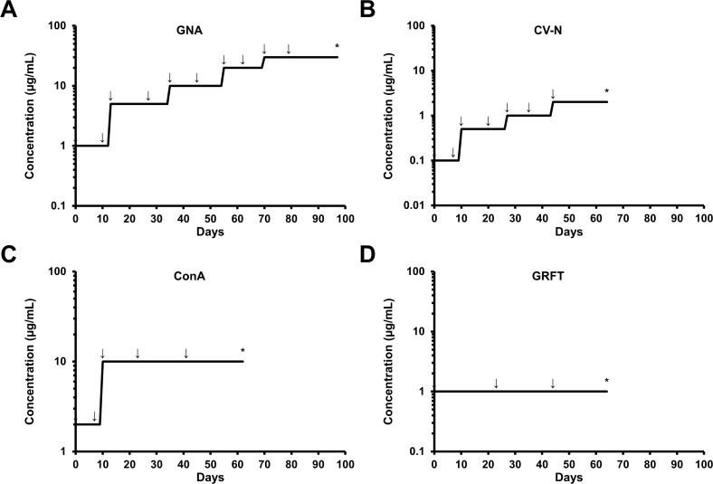
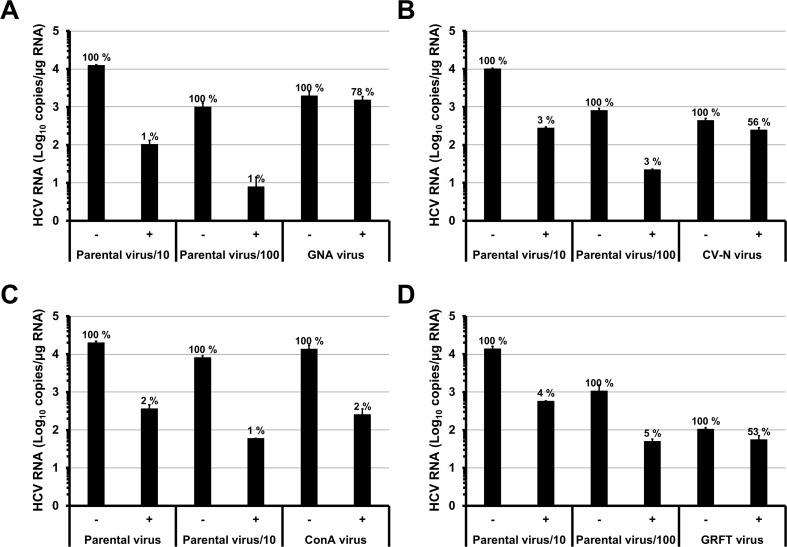
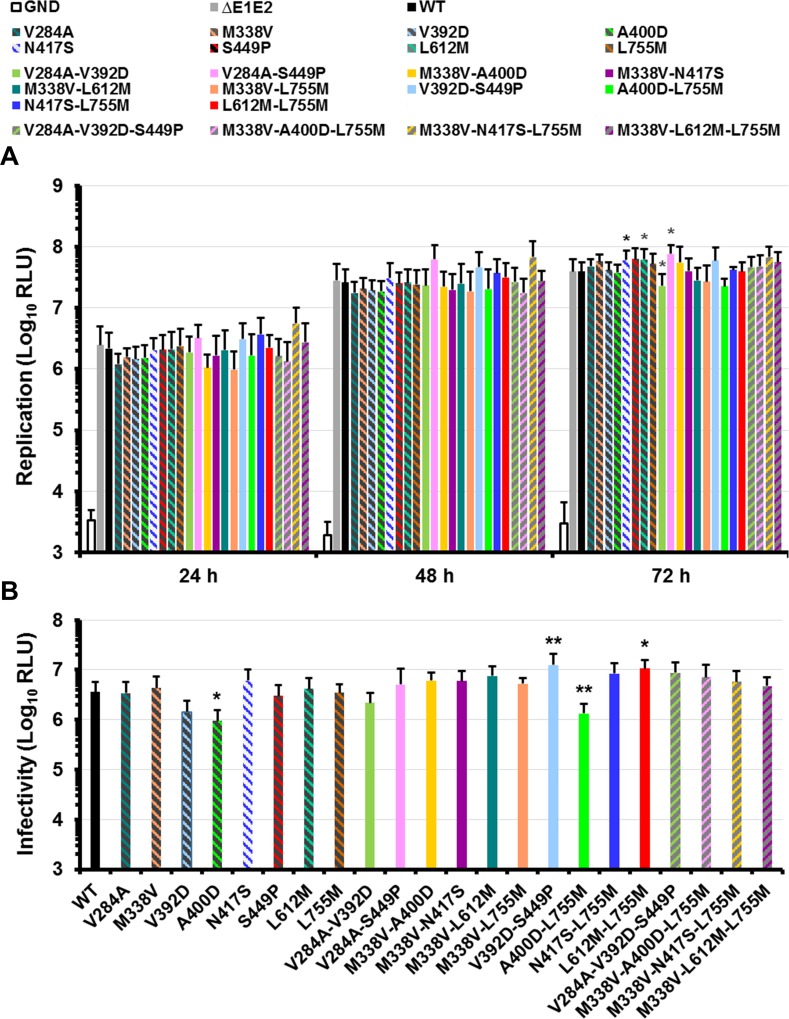
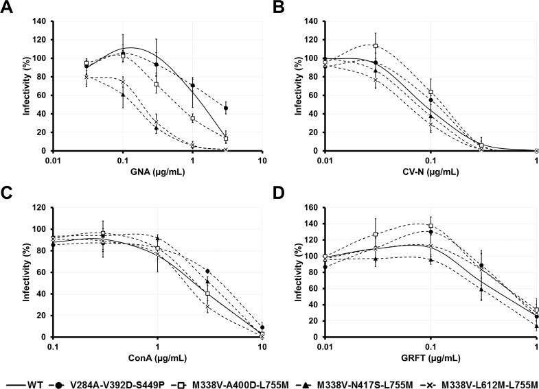
Carbohydrate binding agents (CBAs), including natural lectins, are more and more considered as broad-spectrum antivirals. These molecules are able to directly inhibit many viruses such as Human Immunodeficiency Virus (HIV), Hepatitis C Virus (HCV), Dengue Virus, Ebola Virus or Severe Acute Respiratory Syndrome Coronavirus through binding to envelope protein N-glycans. In the case of HIV, it has been shown that CBAs select for mutant viruses with N-glycosylation site deletions which are more sensitive to neutralizing antibodies. In this study we aimed at evaluating the HCV resistance to CBAs in vitro. HCV was cultivated in the presence of increasing Galanthus nivalis agglutinin (GNA), Cyanovirin-N, Concanavalin-A or Griffithsin concentrations, during more than eight weeks. At the end of lectin exposure, the genome of the isolated strains was sequenced and several potential resistance mutations in the E1E2 envelope glycoproteins were identified. The effect of these mutations on viral fitness as well as on sensitivity to inhibition by lectins, soluble CD81 or the 3/11 neutralizing antibody was assessed. Surprisingly, none of these mutations, alone or in combination, conferred resistance to CBAs. In contrast, we observed that some mutants were more sensitive to 3/11 or CD81-LEL inhibition. Additionally, several mutations were identified in the Core and the non-structural proteins. Thus, our results suggest that in contrast to HIV, HCV resistance to CBAs is not directly conferred by mutations in the envelope protein genes but could occur through an indirect mechanism involving mutations in other viral proteins. Further investigations are needed to completely elucidate the underlying mechanisms.


Conflict of interest statement

Competing Interests: The authors have declared that no competing interests exist.

Figures

Fig 1
Fig 1. Schedule of drug-escalating selection of GNA, CV-N, ConA and GRFT resistant HCV strains as a function of time.
HuH-7-RFP-NLS-IPS cells were infected in the presence of GNA (A), CV-N (B), ConA (C) or GRFT (D). Infected cells were subcultured every three to four days in the presence of the lectins. When 100% cells were infected (indicated by arrows), supernatants were recovered and used to infect naive cells in the presence of the lectins. The concentrations of each lectin were increased in a stepwise manner as indicated. Stars indicate the time points when the virus isolates were recovered and sequenced.
Fig 2
Fig 2. Sensitivity of the strains isolated after selection to inhibition by lectins.
HuH-7-RFP-NLS-IPS cells were inoculated with the strains isolated after selection (GNA, CV-N, ConA and GRFT virus) or different dilutions of the parental strain, in the absence (-) or presence (+) of 3 μg/mL of GNA (A), 0.3 μg/mL of CV-N (B), 10 μg/mL of ConA (C) or 1 μg/mL of GRFT (D). After 48 h, cells were lysed and intracellular HCV RNA were quantified by RT-qPCR. Results are expressed as means ± S.D. of duplicates and percentages are indicated.
Fig 3
Fig 3. Effect of the E1E2p7 mutations on viral genome replication and infectious virus production.
(A) HuH-7-RFP-NLS-IPS cells were transfected with WT or mutated HCV genomes. An assembly-deficient virus (ΔE1E2) and a replication-defective virus (GND) were used as controls. Replication was assessed at 4, 24, 48 and 72 h by measuring Renilla Luciferase activities in transfected cells. Results are expressed as relative light units (RLU) normalized at 4 h and are reported as the means ± standard deviation (S.D.) of at least three independent experiments. (B) The supernatants of transfected cells were recovered at 48 h and incubated for 4 h with naive HuH-7-RFP-NLS-IPS cells. Luciferase assays were performed on infected cells at 72 h post-infection. Results are normalized by the replication at 48 h, expressed as RLU and are reported as the means ± S.D. of at least three independent experiments. (*: p < 0.05; **: p < 0.001). Luciferase activities obtained with ΔE1E2 supernatants were around 103 RLU.
Fig 4
Fig 4. Effect of the E1E2p7 mutations on E1E2 glycoprotein expression.
HuH-7-RFP-NLS-IPS cells were transfected with WT or mutated HCV genomes. An assembly-deficient virus (ΔE1E2) was used as control. Expression of E1E2 viral glycoproteins and actin was analyzed in cell lysates 72h post-electroporation by western blotting with specific MAbs (A4 [anti-E1], 3/11 [anti-E2] and C4 [anti-actin]).
Fig 5
Fig 5. Effect of the E1E2p7 mutations on sensitivity to inhibition by GNA, CV-N, ConA and GRFT.
Inhibition assays were performed by incubating WT or mutant HCVcc with various concentrations of GNA (A), CV-N (B), ConA (C) or GRFT (D). After a 1 h incubation at 37°C, mixes were put into contact with target cells for 4 h. Luciferase assays were performed on infected cells at 72 h post-infection. Results are expressed as percentages of infectivity compared to infection in absence of inhibitory protein and are reported as the means ± S.D. of at least three independent experiments.
Fig 6
Fig 6. Effect of the E1E2p7 mutations on sensitivity to inhibition by MAb 3/11 and CD81-LEL.
Inhibition assays were performed by incubating WT or mutant HCVcc with various concentrations of MAb 3/11 (A) or CD81-LEL (B). After a 1 h incubation at 37°C, mixes were put into contact with target cells for 4 h. Luciferase assays were performed on infected cells at 72 h post-infection. Results are expressed as percentages of infectivity compared to infection in absence of inhibitory protein and are reported as the means ± S.D. of at least three independent experiments.

References

    1. Simmonds P. The origin of hepatitis C virus. Curr Top Microbiol Immunol. 2013;369:1–15. Epub 2013/03/07. 10.1007/978-3-642-27340-7_1 . - DOI - PubMed
    1. Pawlotsky JM, Feld JJ, Zeuzem S, Hoofnagle JH. From non-A, non-B hepatitis to hepatitis C virus cure. J Hepatol. 2015;62(1S):S87–S99. Epub 2015/04/29. S0168-8278(15)00080-X [pii] 10.1016/j.jhep.2015.02.006 . - DOI - PubMed
    1. Dubuisson J, Cosset FL. Virology and cell biology of the hepatitis C virus life cycle—an update. J Hepatol. 2014;61(1 Suppl):S3–S13. Epub 2014/12/03. 10.1016/j.jhep.2014.06.031 S0168-8278(14)00461-9 [pii]. . - DOI - PubMed
    1. Vieyres G, Thomas X, Descamps V, Duverlie G, Patel AH, Dubuisson J. Characterization of the envelope glycoproteins associated with infectious hepatitis C virus. J Virol. 2010;84(19):10159–68. Epub 2010/07/30. JVI.01180-10 [pii] 10.1128/JVI.01180-10 - DOI - PMC - PubMed
    1. Falkowska E, Kajumo F, Garcia E, Reinus J, Dragic T. Hepatitis C virus envelope glycoprotein E2 glycans modulate entry, CD81 binding and neutralization. J Virol. 2007;81(15):8072–9. . - PMC - PubMed

Publication types