. 2016 Mar 22;7(12):13651-66.
doi: 10.18632/oncotarget.7296.

Hyperinsulinemia enhances interleukin-17-induced inflammation to promote prostate cancer development in obese mice through inhibiting glycogen synthase kinase 3-mediated phosphorylation and degradation of interleukin-17 receptor

Affiliations

Hyperinsulinemia enhances interleukin-17-induced inflammation to promote prostate cancer development in obese mice through inhibiting glycogen synthase kinase 3-mediated phosphorylation and degradation of interleukin-17 receptor

Sen Liu et al. Oncotarget. .

Abstract

Interleukin-17 (IL-17) plays important roles in inflammation, autoimmune diseases, and some cancers. Obese people are in a chronic inflammatory state with increased serum levels of IL-17, insulin, and insulin-like growth factor 1 (IGF1). How these factors contribute to the chronic inflammatory status that promotes development of aggressive prostate cancer in obese men is largely unknown. We found that, in obese mice, hyperinsulinemia enhanced IL-17-induced expression of downstream proinflammatory genes with increased levels of IL-17 receptor A (IL-17RA), resulting in development of more invasive prostate cancer. Glycogen synthase kinase 3 (GSK3) constitutively bound to and phosphorylated IL-17RA at T780, leading to ubiquitination and proteasome-mediated degradation of IL-17RA, thus inhibiting IL-17-mediated inflammation. IL-17RA phosphorylation was reduced, while the IL-17RA levels were increased in the proliferative human prostate cancer cells compared to the normal cells. Insulin and IGF1 enhanced IL-17-induced inflammatory responses through suppressing GSK3, which was shown in the cultured cell lines in vitro and obese mouse models of prostate cancer in vivo. These findings reveal a mechanism underlying the intensified inflammation in obesity and obesity-associated development of aggressive prostate cancer, suggesting that targeting GSK3 may be a potential therapeutic approach to suppress IL-17-mediated inflammation in the prevention and treatment of prostate cancer, particularly in obese men.

Keywords: GSK3; hyperinsulinemia; interleukin-17; obesity; prostate cancer.


Conflict of interest statement

CONFLICTS OF INTERESTS

The authors declared no conflicts of interests.

Figures
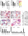
Figure 1
Figure 1. High-fat diet-induced obesity promotes prostate cancer formation in Pten conditional knockout mouse prostates
(A) Mouse body weight at age of 30 weeks. *P < 0.001 (Student's t test). (B) The tissue weight of inguinal fat and epididymal fat. *P < 0.001 (Student's t test). (C) The genitourinary (GU) block weight. *P < 0.01 (Student's t test). (D) Percentages of PIN and invasive cancer in dorsal, lateral, and ventral prostatic lobes. *P < 0.001 (Kruskal–Wallis test). (E) Representatives of H & E-stained dorsal prostatic lobes showing mouse prostatic intraepithelial neoplasia (PIN) and invasive (or microinvasive) adenocarcinomas; the red windows highlight the selected regions at x 400 magnification; scale bar, 100 μm. (F) Western blot analysis of the proteins isolated from the representative prostate tissues of obese and lean mice at age of 30 weeks. Anti-P-GSK3α antibodies recognize Ser21 phosphorylation site and anti-P-GSK3β antibodies recognize Ser9 phosphorylation site. (G,H) qRT-PCR analysis of mRNA expression in lean and obese mouse prostates. *P < 0.05 (one-way ANOVA). Data represent mean ± standard deviation of 10 mice per group (n = 10).
Figure 2
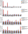
Figure 2. Induction of gene expression in Gsk3β+/+ (wild-type) and Gsk3β−/− (knockout) mouse embryonic fibroblasts (MEFs) by IL-17 family cytokines
(AD) MEFs were treated with 20 ng/ml mouse recombinant IL-17A (A), IL-17F (B), IL-17C (C), and IL-17E (D) for 2 h. Gene expression was determined using qRT-PCR analysis. The levels of the control group (treated with phosphate-buffered saline) were taken as the basal levels. Data represent the mean ± standard deviation of three independent experiments (n = 3); *P < 0.05 (Student's t test).
Figure 3
Figure 3. GSK3β binds to and phosphorylates IL-17RA
(AD) 293 cells were co-transfected with Flag-IL-17RA, V5-IL-17RC, and HA-Act1, labeled with 32P orthophosphate, and treated with 20 ng/ml IL-17A for 20 min; IP with anti-HA; autoradiography (A) followed by IB (B-D). (E) Anti-Flag IP followed by IB. (FG) Co-IP of Flag-IL-17RA and HA-GSK3β. (H) 293 cells were co-transfected with Flag-IL-17RA, WT HA-GSK3β or kinase-dead mutant HA-GSK3βK85A, labeled with 32P orthophosphate, or treated with 20 mM LiCl for 2 h; autoradiography (32P) followed by IB. WE, whole cell extract.
Figure 4
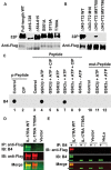
Figure 4. GSK3β phosphorylates IL-17RA at T780
(AB) 293 cells were transfected with Flag-IL-17RA and its mutants and labeled with 32P orthophosphate; autoradiography (32P) followed by IB. (C) IL-17RA peptide phosphorylated at T780 (p-Peptide) was treated with CIP, while IL-17RA peptide with wild-type T780 (Peptide) or with mutant T780A (mut-Peptide) were treated with recombinant GSK3, followed with dot blot analysis using B4 antibodies. (DE) Flag-IL-17RA, Flag-IL-17RAT780A mutant, or empty vector was transfected into 293 cells; HeLa cells were not transfected; IP with anti-Flag, B4 (+), or control IgG (−), followed by IB; *indicates endogenous P-IL-17RA.
Figure 5
Figure 5. AZD5363 antagonizes insulin-induced enhancement of IL-17 responses through restoring IL-17RA phosphorylation
(A) and (D) 239 cells stably expressing Flag-IL-17RA (293-IL-17RA cell line) and HeLa cells were treated with 2 μM AZD5363 (a pan-Akt inhibitor) and/or 50 ng/ml insulin for the indicated time periods; Western blot analysis was performed for the indicated proteins; exogenous, IL-17RA transfected into 293 cells; endogenous, endogenous IL-17RA expressed in 293 and HeLa cells. (B) and (C) 293-IL-17RA cells were treated with 20 ng/ml IL-17A, with or without 2 μM AZD5363 and/or 50 ng/ml insulin for 2 h; IL-17-downstream gene expression was determined using qRT-PCR analysis; *P < 0.05 compared to each single treatment group; **P < 0.05 compared to the combined insulin and IL-17 treatment group (one-way ANOVA). (E) and (F) HeLa cells were treated with 20 ng/ml IL-17A, with or without 2 μM AZD5363 and/or 50 ng/ml insulin for 2 h; IL-17-downstream gene expression was determined using qRT-PCR analysis; *P < 0.05 compared to each single treatment group; **P < 0.05 compared to the combined insulin and IL-17 treatment group (one-way ANOVA). Data represent the mean ± standard deviation of three independent experiments (n = 3).
Figure 6
Figure 6. Phosphorylated IL-17RA is ubiquitinated and degraded by proteasome-mediated mechanism
(AD) 293 cells stably expressing Flag-IL-17RA were treated with 20 mM LiCl or 10 μM MG132, and/or 50 μg/ml CHX; P-IL-17RA was detected using B4 and total exogenous IL-17RA was detected using anti-Flag; the levels of total exogenous IL-17RA (C) were normalized by the levels of GAPDH (D); exo, exogenously transfected IL-17RA; endo, endogenously expressed IL-17RA; *P < 0.05 compared to LiCl and MG132 treated groups (Student's t test). (E) Flag-IL-17RA and Flag-IL-17RA-T780A mutant were co-transfected with HA-tagged ubiquitin (HA-Ub), with or without 10 μM MG132 treatment; non-specific, an unknown band detected by anti-HA antibodies, which was not IL-17RA or IgG heavy chain (see Supplementary Figure S3C). (F) Flag-IL-17RA and Flag-IL-17RA-T780A mutant were co-transfected with HA-tagged ubiquitin (HA-Ub) or K48-only ubiquitin (HA-K48), with 10 μM MG132 treatment, followed by IP and IB.
Figure 7
Figure 7. Phosphorylation of IL-17RA is detected in human prostate tissues
(AI) Human normal prostate, PIN, and prostate cancer tissues were double stained for P-IL-17RA using B4 (arrowheads) and for basal cells using anti-p63 (arrows). (J) Quantification of P-IL-17RA staining.

References

    1. Moseley TA, Haudenschild DR, Rose L, Reddi AH. Interleukin-17 family and IL-17 receptors. Cytokine Growth Factor Rev. 2003;14:155–74. - PubMed
    1. Gaffen SL, Jain R, Garg AV, Cua DJ. The IL-23-IL-17 immune axis: from mechanisms to therapeutic testing. Nat Rev Immunol. 2014;14:585–600. - PMC - PubMed
    1. Shi Y, Ullrich SJ, Zhang J, Connolly K, Grzegorzewski KJ, Barber MC, Wang W, Wathen K, Hodge V, Fisher CL, Olsen H, Ruben SM, Knyazev I, et al. A novel cytokine receptor-ligand pair. Identification, molecular characterization, and in vivo immunomodulatory activity. J Biol Chem. 2000;275:19167–76. - PubMed
    1. Reynolds JM, Lee YH, Shi Y, Wang X, Angkasekwinai P, Nallaparaju KC, Flaherty S, Chang SH, Watarai H, Dong C. Interleukin-17B Antagonizes Interleukin-25-Mediated Mucosal Inflammation. Immunity. 2015;42:692–703. - PMC - PubMed
    1. McAleer JP, Kolls JK. Directing traffic: IL-17 and IL-22 coordinate pulmonary immune defense. Immunol Rev. 2014;260:129–44. - PMC - PubMed

MeSH terms