. 2016 May:54:233-242.
doi: 10.1016/j.bbi.2016.02.007. Epub 2016 Feb 9.

Neutrophil depletion after subarachnoid hemorrhage improves memory via NMDA receptors

Affiliations

Neutrophil depletion after subarachnoid hemorrhage improves memory via NMDA receptors

Jose Javier Provencio et al. Brain Behav Immun. 2016 May.

Abstract

Cognitive deficits after aneurysmal subarachnoid hemorrhage (SAH) are common and disabling. Patients who experience delayed deterioration associated with vasospasm are likely to have cognitive deficits, particularly problems with executive function, verbal and spatial memory. Here, we report neurophysiological and pathological mechanisms underlying behavioral deficits in a murine model of SAH. On tests of spatial memory, animals with SAH performed worse than sham animals in the first week and one month after SAH suggesting a prolonged injury. Between three and six days after experimental hemorrhage, mice demonstrated loss of late long-term potentiation (L-LTP) due to dysfunction of the NMDA receptor. Suppression of innate immune cell activation prevents delayed vasospasm after murine SAH. We therefore explored the role of neutrophil-mediated innate inflammation on memory deficits after SAH. Depletion of neutrophils three days after SAH mitigates tissue inflammation, reverses cerebral vasoconstriction in the middle cerebral artery, and rescues L-LTP dysfunction at day 6. Spatial memory deficits in both the short and long-term are improved and associated with a shift of NMDA receptor subunit composition toward a memory sparing phenotype. This work supports further investigating suppression of innate immunity after SAH as a target for preventative therapies in SAH.

Keywords: Cerebral vasospasm; Delayed neurological deterioration after subarachnoid hemorrhage; Innate inflammation; Memory deficits; Neutrophils; Subarachnoid hemorrhage.


Conflict of interest statement

Conflict of Interest Statement: The authors have declared that no conflict of interest exists.

Figures

Figure 1
Figure 1. Subarachnoid hemorrhage leads to long term spatial memory loss due to NMDA receptor mediated abnormalities in late long-term potentiation (L-LTP) that occur between the third and sixth day
Barnes maze (BMT) and Morris water maze (MWM) analysis completed on the same cohort of animals over the first 9 days (BMT) and one month after (MWM) SAH or Sham. (A) There was significantly worse performance on BMT in animals in the SAH group. (B) This was consistent in the MWM performed at one month after SAH/Sham suggesting that the spatial memory deficits after SAH are present early and sustained as late as a month. (C) The diagram shows the experimental design of the electrophysiology experiments performed with stimulation of Schaffer collateral fibers between the CA3 region of the hippocampus and the CA1 region with acquisition of data from synapses between Shaffer collateral axons and pyramidal cell bodies. (D) The EPSP slope vs. time graph shows significantly preserved L-LTP on day 3 in SAH animals compared to Sham (n=3 animals/group with 7 slices). (E) At day 6, SAH animals lose L-LTP while Sham do not (n=3 animals/group with 9 slices). (F) Isolation of the NMDA receptor using bath conditions shown in the figure reveal continued loss of L-LTP suggesting the NMDAR (and not the AMPAR) is responsible for the effect (n=3 animals/group with 6 total slices). (G) shows a graphical display of the difference in L-LTP between SAH and sham at day 3, 6 and at day 6 with NMDA isolation.
Figure 2
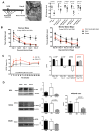
Figure 2. Neutrophil depletion three days after SAH Improves vasospasm and spatial memory task performance through changes in the proportion of NMDA subunit composition
(A) Neutrophil depletion using the 1A8 antibody prior to SAH does not significantly improve vasospasm measured 1mm from the carotid terminus in the middle cerebral artery 6 days after SAH (unpaired Student’s t-test). The bar on the left shows the experimental design and a picture of the measurement point on the middle cerebral artery. Depleting neutrophils 12 hours (0.5 days) and 1 day after SAH also does not significantly improve vasospasm (retained significant difference between SAH and Sham) (unpaired Student’s t-test). Conversely, depletion of neutrophils 3 days after SAH does improve vasospasm. (B) Mice depleted of neutrophils 3 days after SAH (red line) show significant improvement in Barnes Maze test (BMT) and Morris Water maze (MWM) performance compared to SAH. (C) The EPSP slope vs. time graph shows preserved L-LTP on day 6 in SAH animals compared to Sham (n=3 animals/group). The bar graph adds the neutrophil depletion to the Sham/SAH data presented in Figure 1 and shows no significant difference in L-LTP between SAH and Sham animals. (D) Western blot analysis of NR1, NR2A and NR2B NMDAR subunits in hippocampal cell membrane fractions show that NR1, NR2A and NR2B are not significantly altered after SAH but NR2A is significantly decreased after depletion of neutrophils 3 days after SAH. Likewise, the NR2A/NR2B ratio is also significantly decreased after neutrophil depletion.
Figure 3
Figure 3. Microglial activation is increased after SAH but few neutrophils enter the brain
(A) Neutrophil staining with the Ly6G antibody 1A8 (TO-Pro-3 stains nuclei) shows no neutrophils in the area of the hippocampus on day 1, 3 or 6 or SAH. For comparison, naïve spinal cord (SC), SC at onset after EAE (experimental autoimmune encephalitis, an experimental model of spinal cord inflammation and demyelination) or at peak EAE: the later two showing neutrophils in the parenchyma (arrows). White bar represents 200 μm and yellow bar represents 25 μm. (B) In a LysM-EGFP mouse (Lyz2tm1.1Graf), we compared SAH and sham mice. In the SAH mice, we found few Anti-GFP+, Iba-1-, cells that represent non-macrophage perivascular infiltrating myeloid cells in the hippocampus. (C) Microglial staining using Iba-1 (TO-Pro-3 nuclei) shows increased size and decreased ramifications six days after SAH (at the same time as changes in behavior). Depletion of neutrophils increases microglial ramification and decreases the number of Iba-1 positive cells. Solid bars represent 250 μm and dotted lines represent 50 μm. The graph shows total fluorescence intensity of Iba-1 positive calls in the hippocampus 6 days after SAH.

References

    1. van Gijn J, Rinkel GJ. Subarachnoid haemorrhage: diagnosis, causes and management. Brain. 2001;124(Pt 2):249–278. - PubMed
    1. Macdonald RL, Higashida RT, Keller E, et al. Clazosentan, an endothelin receptor antagonist, in patients with aneurysmal subarachnoid haemorrhage undergoing surgical clipping: a randomised, double-blind, placebo-controlled phase 3 trial (CONSCIOUS-2) Lancet Neurol. 2011;10(7):618–625. - PubMed
    1. Hop JW, Rinkel GJ, Algra A, van Gijn J. Changes in functional outcome and quality of life in patients and caregivers after aneurysmal subarachnoid hemorrhage.[see comment] Journal of Neurosurgery. 2001;95(6):957–963. - PubMed
    1. Barker FG, 2nd, Ogilvy CS. Efficacy of prophylactic nimodipine for delayed ischemic deficit after subarachnoid hemorrhage: a metaanalysis. J Neurosurg. 1996;84(3):405–414. - PubMed
    1. Frontera JA, Fernandez A, Schmidt JM, et al. Defining vasospasm after subarachnoid hemorrhage: what is the most clinically relevant definition? Stroke. 2009;40(6):1963–1968. - PubMed

Publication types

Substances