

Role of hypoxia during nephrogenesis
- PMID: 26872484
- PMCID: PMC4982845
- DOI: 10.1007/s00467-016-3333-5
Role of hypoxia during nephrogenesis
Abstract
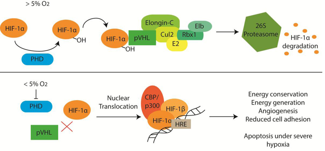
Mammals develop in a physiologically hypoxic state, and the oxygen tension of different tissues in the embryo is precisely controlled. Deviation from normal oxygenation, such as what occurs in placental insufficiency, can disrupt fetal development. Several studies demonstrate that intrauterine hypoxia has a negative effect on kidney development. As nascent nephrons are forming from nephron progenitors in the nephrogenic zone, they are exposed to varying oxygen tension by virtue of the development of the renal vasculature. Thus, nephrogenesis may be linked to oxygen tension. However, the mechanism(s) by which this occurs remains unclear. This review focuses on what is known about molecular mechanisms active in physiological and pathological hypoxia and their effects on kidney development.
Keywords: Hypoxia; Nephrogenesis; Nephron progenitors; Placental insufficiency.
Conflict of interest statement
The authors declare no conflict of interest.
Figures
References
-
- Okazaki K, Maltepe E. Oxygen, epigenetics and stem cell fate. Regen Med. 2006;1:71–83. - PubMed
-
- Ivanovic Z. Hypoxia or in situ normoxia: The stem cell paradigm. J Cell Physiol. 2009;219:271–275. - PubMed
-
- Mohyeldin A, Garzon-Muvdi T, Quinones-Hinojosa A. Oxygen in stem cell biology: a critical component of the stem cell niche. Cell Stem Cell. 2010;7:150–161. - PubMed
-
- Dunwoodie SL. The role of hypoxia in development of the Mammalian embryo. Dev Cell. 2009;17:755–773. - PubMed
Publication types
MeSH terms
Grants and funding
LinkOut - more resources
Full Text Sources
Other Literature Sources
Research Materials
