. 2016 Feb 16;44(2):246-58.
doi: 10.1016/j.immuni.2016.01.008. Epub 2016 Feb 9.

The Wnt Antagonist Dickkopf-1 Promotes Pathological Type 2 Cell-Mediated Inflammation

Affiliations

The Wnt Antagonist Dickkopf-1 Promotes Pathological Type 2 Cell-Mediated Inflammation

Wook-Jin Chae et al. Immunity. .

Abstract

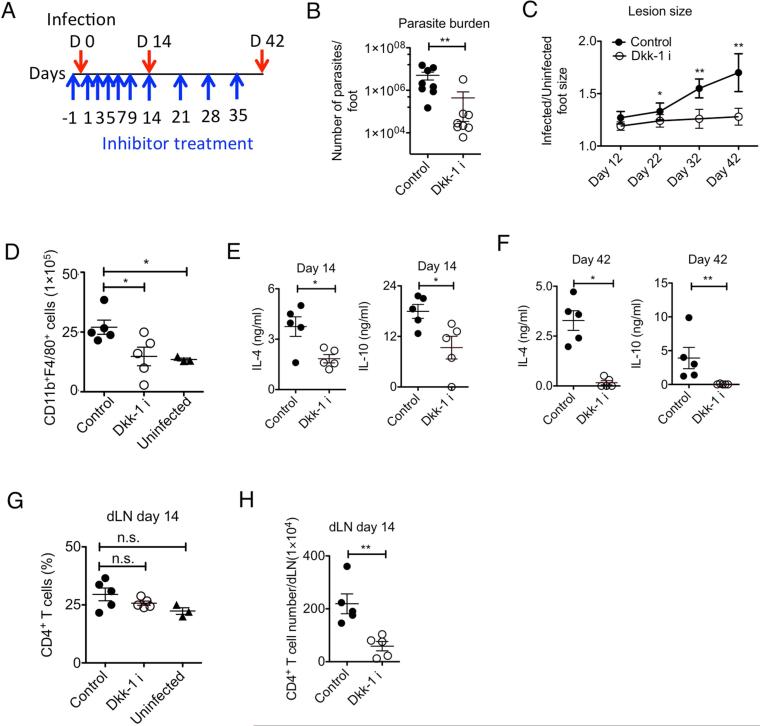
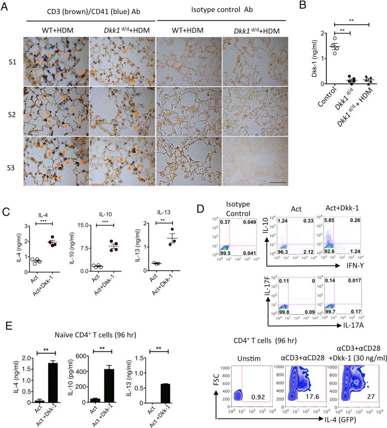
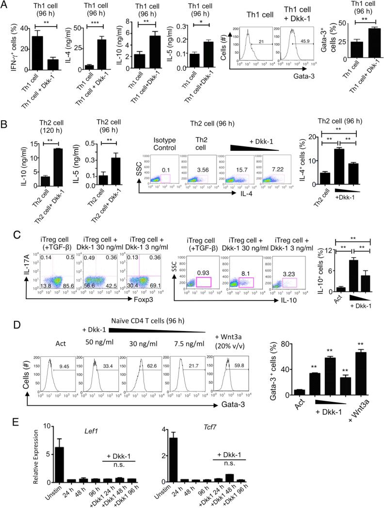
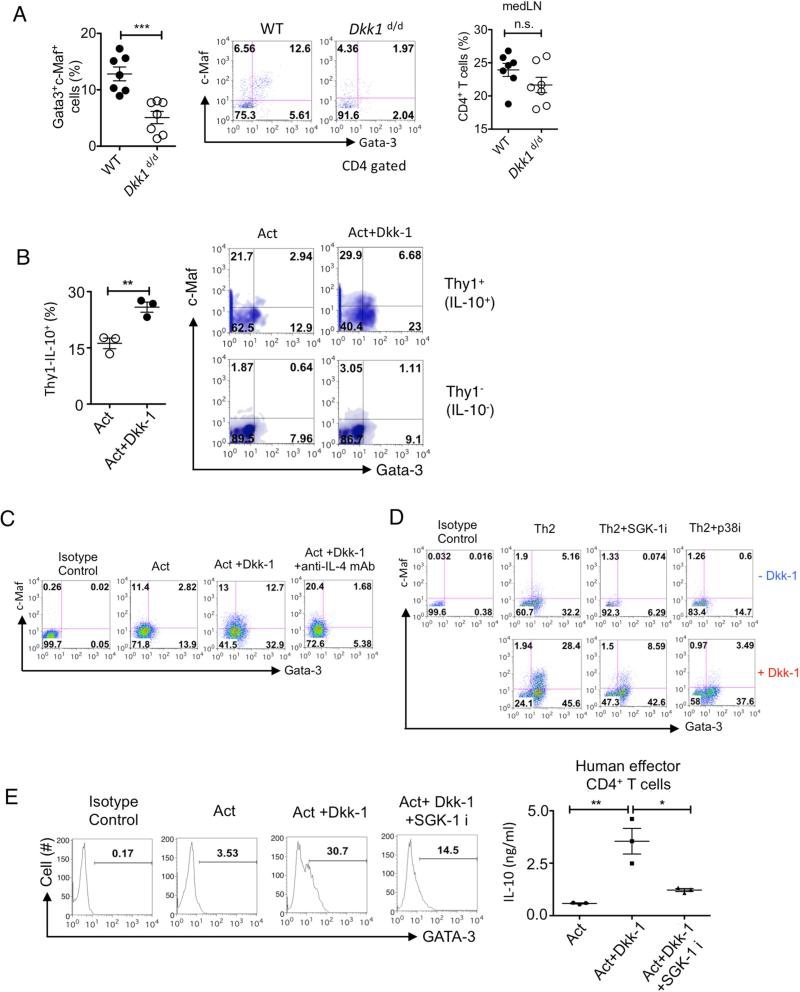
Exposure to a plethora of environmental challenges commonly triggers pathological type 2 cell-mediated inflammation. Here we report the pathological role of the Wnt antagonist Dickkopf-1 (Dkk-1) upon allergen challenge or non-healing parasitic infection. The increased circulating amounts of Dkk-1 polarized T cells to T helper 2 (Th2) cells, stimulating a marked simultaneous induction of the transcription factors c-Maf and Gata-3, mediated by the kinases p38 MAPK and SGK-1, resulting in Th2 cell cytokine production. Circulating Dkk-1 was primarily from platelets, and the increase of Dkk-1 resulted in formation of leukocyte-platelet aggregates (LPA) that facilitated leukocyte infiltration to the affected tissue. Functional inhibition of Dkk-1 impaired Th2 cell cytokine production and leukocyte infiltration, protecting mice from house dust mite (HDM)-induced asthma or Leishmania major infection. These results highlight that Dkk-1 from thrombocytes is an important regulator of leukocyte infiltration and polarization of immune responses in pathological type 2 cell-mediated inflammation.


Figures

Figure 1
Figure 1. Reduced expression of Dkk-1 protects the host from house dust mite (HDM)-induced asthma
(A) Circulating concentrations of Dkk-1 in eight-week-old female Dkk1d/d mice (n=5) and their wildtype (WT) littermate control mice (n=5) (B) A scheme of HDM challenge protocol (10 μg/mouse/time) (C, D) nine to twelve-week old Dkk1d/d (n=7) and their littermate controls (n=7) were analyzed. Total leukocytes (CD45+), neutrophils eosinophils from BAL fluid (BALF) and lung tissue homogenates were quantitated. (E) CD4+ T cells from lung tissue homogenates were quantitated. (F) Supernatants from mediastinal lymph node cells that were stimulated with HDM extract for 4 days were analyzed by ELISA. (G,H,I) Lung tissues from each mouse were scored after H&E and PAS staining. For F, G, and H, controls were challenged with PBS (n=3), and One-Way ANOVA analysis with Dunnet's post-hoc test was performed. Original magnification is 10×. Scale bar in lower right panel represents 10 μm. Small horizontal lines indicate the mean (± s.e.m.). A representative of two independent experiments is shown. Student's t-test was performed. ***, p<0.0005, **, p<0.005, *, p<0.05. See also supplementary Figures 1 and 2.
Figure 2
Figure 2. Functional inhibition of Dkk-1 protects the host from chronic inflammation caused by L. major
(A) A schematic diagram for Dkk-1 inhibitor treatment (10 μg/kg/time) in six-week-old female BALB/c mice. (B) The infected foot from each mouse was analyzed for parasite burden at day 42 and (C) Lesion size was measured (n=8/group). (D) Two weeks after vehicle (n=5) Dkk-1 inhibitor treatment (n=5) based on (A), macrophages in the infected hindfoot were counted by flow cytometry and compared with infected (untreated) and uninfected BALB/c mice (n=3). (E, F) Two weeks and 6 weeks after infection, draining lymph node cells from each mouse (n=5/group) were stimulated with sLMAG. (G, H) CD4 T cell numbers and percentage in draining lymph nodes from each group was determined by flow cytometry. Small horizontal lines indicate the mean (± s.e.m.). A representative of two independent experiments is shown. Student's t-test (B, C, E-H) and One-Way-ANOVA analysis with Dunnet's post-hoc test (D,G) were performed. **, p<0.005, *, p<0.05.
Figure 3
Figure 3. Dkk-1 induces Th2 cell polarization
(A) Dkk1d/d mice and their wildtype littermate controls were challenged with HDM extract allergen shown in Figure 1B. Mice were sacrificed and lungs were harvested for immunohistochemistry for CD3 (dark brown), CD41 (blue) and isotype control antibodies. Orange arrowheads in the images indicate co-localized CD3+ T cells with CD41+ platelets. S1, S2, and S3 indicate individual animal in each group. Original magnification is 63×. Scale bar in lower right panel represents 10 μm. (B) Dkk1d/d mice (n=5) were challenged with 50 μg HDM extract per mouse and plasma samples were collected 24 hours later for ELISA. (C, D) Splenic CD4 T cells from 6-8 week-old C57BL/6 mice were activated with anti-CD3 and anti-CD28 antibodies (Act) with or without Dkk-1 (30 ng/ml unless indicated) for 4 days. Supernatants were analyzed by ELISA (C) and flow cytometry (D). For IL-4 in (D), splenic CD4 T cells from 8 week-old IL-4-GFP reporter mice were used. (E) Naïve CD4+ T cells were stimulated anti-CD3 and anti-CD28 antibodies for 96 hr with or without Dkk-1 for flow cytometry analysis and ELISA. Small horizontal lines and error bars indicate the mean (± s.e.m.). A representative of three independent experiments is shown. Student's t-test was performed. ***, p < 0.0001, **, p< 0.005, *, p<0.05.
Figure 4
Figure 4. Dkk-1 induces Th2 cytokines in various T cell differentiation conditions
(A,B) Naïve CD4 T cells were stimulated under Th1 (A), Th2 (B) polarization conditions for 96 hours with or without Dkk-1 for flow cytometry analysis and ELISA. (C) Splenic naïve CD4+ T cells were isolated and differentiated into iTreg for 5 days with 100 U/ml IL-2 and 1.5 ng/ml TGF-β with or without 30 ng/ml and 3 ng/ml of Dkk-1 was treated during the culture. Foxp3 and IL-17A expression (left panels) and IL-10 expression (right panels) were measured by flow cytometry. (D) Splenic naïve CD4+ T cells from 8 week-old C57BL/6 mice were treated with Dkk-1 with varying doses of Dkk-1 (50, 30, 7.5 ng/ml) or Wnt3a (20% v/v) for 96 h in the presence of anti-CD3 and anti-CD28 antibody stimulation to measure Gata-3 expression amounts. (E) Splenic naïve CD4+ T cells from 8 week-old C57BL/6 mice were stimulated with or without Dkk-1 (30 ng/ml) for 96 hr. RNA was harvested and analyzed by real time qPCR. All results were statistically non-significant. All experiments are a representative of two to three independent experiments. For (A), student's t-test was performed. For (B-E), One way-ANOVA analysis with Bonferonni's post-hoc test was performed. Small horizontal lines and error bars indicate the mean (± s.e.m.). ***, p < 0.0001, **, p< 0.005, *, p<0.05..
Figure 5
Figure 5. Dkk-1 directs TH2 cell polarization by p38 MAPK and SGK-1 to induce c-Maf and Gata-3
(A) Mediastinal lymph node cells from Figure 1D were stimulated with HDM extract for 4 days and the percentages of Gata-3+ and c-Maf+ in CD4+ T cells were quantitated by flow cytometry. (B) Splenic CD4+ T cells from 12-week old Thy1-IL-10 reporter mice were activated with or without Dkk-1 (30 ng/ml) for 84 hr. Cells were gated for Thy1+ and Thy1. (C) Splenic CD4+ T cells were activated with or without Dkk-1 or anti-IL-4 mAb (10 μg/ml) for 4 days and analyzed by flow cytometry. (D) Naïve CD4+ T cells were stimulated under Th2 polarization conditions for 96 hr with or without Dkk-1 for flow cytometry analysis. SGK-1 inhibitor (2 μM) or p38 inhibitor (10 μM) was added at 0 h. (E) PBMCs from healthy volunteers were used to isolate human CD4+ T cells, and then stimulated with human anti-CD3 mAb (OKT3) and anti-CD28 mAb for 4 days with or without Dkk-1 (30 ng/ml) and SGK-1 inhibitor GSK653094 (2 μM). Cells were analyzed by flow cytometry. Supernatants were harvested and analyzed by ELISA (right panel). A representative of three independent experiments is shown. Student's t-test was performed. ***, p < 0.0001, **, p< 0.005, *, p<0.05, n.s., not significant. For (E), One-way ANOVA analysis with Bonferroni's post-hoc test was performed. See also supplementary Figure 3.
Figure 6
Figure 6. Circulating Dkk-1 is from platelets upon allergen challenge or parasitic infection
(A) Female C57BL/6 mice were challenged with PBS (n=3) or HDM extract (10 μg/challenge/mouse, n=5) as in Figure 1B. (B) Eight-week old female C57BL/6 mice were challenged with HDM extract (30 μg/challenge, n=4) or PBS (n=5) for 4 and 24 hours. Plasma samples were analyzed by ELISA. (C) Dkk1d/d mice (n=7) and their wildtype littermate controls (n=7) were challenged with HDM extract as in Figure 1B. Harvested lungs were analyzed by immunohistochemistry. Original magnification is 10×. Scale bar in lower right panel represents 10 μm. (D) Peripheral blood was collected by cardiac puncture with or without EDTA. (E) Eight-week-old female C57BL/6 mice were injected with platelet depletion antibody (n= 5) or isotype control antibody (n=4). Twenty-four hours later, plasma samples were measured. (F) HDM allergen was challenged as in Figure 1B, and platelets were depleted for 12 hours. (G) Bacterial ds DNA from E.coli (B-ds DNA)(30 μg/mouse,n=4), LPS (3 μg/mouse, n=5), CpG-ODN 1585 (25 μg/mouse, n=4), and House Dust Mite (HDM) allergen extract (50 μg/mouse, n=4) were challenged intranasally in 6 week-old C57BL/6 mice. Twenty-four hours later, plasma samples were collected and Dkk1 concentrations were determined by ELISA. Control (n= 7) mice were given 20 μl of 0.9% NaCl saline. (H) At each time point plasma from female BALB/c mice (6-week old) that were infected with L. major was measured. (I) Platelets were depleted 4 hours prior to parasite infection. Plasma samples from 5-week old female BALB/c mice were analyzed 72 hours after infection. (J) Ten weeks after infection, platelets were depleted for 12 hours in BALB/c mice. (K) Human platelets from four healthy volunteers were activated with sLMAG for 1 hr. S1-S4 designates each healthy donor. X-axis shows dilution of sLMAG in the culture. 1:50 is equivalent to 1×10⁶ parasites. (L) Human platelets (1×10⁸/ml, n=5) were activated with sLMAG (1:50) in the presence of PKCα inhibitor, or PKCβ inhibitor for 1 hour. A representative of two independent experiments is shown. Small horizontal lines indicate the mean (± s.e.m.). Student's t-test (A, D, E), One-way ANOVA with Dunnett's post-hoc test (B, I) and One-way ANOVA with Bonferroni's post-hoc test (F,G,H,J,L) was performed. ***, p< 0.0005, **, p<0.005, *, p<0.05. See also Supplementary Figure 4.
Figure 7
Figure 7. Dkk-1 facilitates leukocyte migration and regulates leukocyte-platelet aggregate formation in type 2 cell-mediated inflammation
(A) Nine-week old Dkk1d/d mice and wildtype littermate controls (n=5-6/group) were challenged with 30 μg HDM allergen extract intranasally. Lung homogenates were quantitated and analyzed by flow cytometry 72 hours after HDM challenge and CD45+ cells were analyzed by flow cytometry. (B) Peripheral blood from nine-week old Dkk1d/d mice and wildtype littermate controls were collected at 4 and 24 hours after allergen challenge, and analyzed for the percentages of CD45+CD41+ cells. (C) Dkk-1 protein (300 ng) was injected intraperitoneally. CD45 cells were analyzed by flow cytometry. NE, neutrophils, EO, eosinophils. (D) Schematic diagram of Dkk-1 inhibitor treatment protocol for each group (n=5 for each group) in HDM-induced asthma model. HDM allergen extract (10 μg) was challenged intranasally at the indicated time points. CD45+ leukocytes numbers in BALF and Lung (E), neutrophils, eosinophils, and CD4 T cell numbers in the lungs (F) and the number of CD4+ T cells in mediastinal LNs were counted (G). After 4 days of stimulation of med LN cells, cytokines were measured by ELISA (H). H&E and PAS staining were scored (I, J). ET, early treatment; LT, late treatment; FT, full treatment; PC, positive control. Small horizontal lines and error bars indicate the mean (± s.e.m.). A representative of two independent experiments is shown. One-way ANOVA analysis with Dunnet's post-hoc test was performed. ***, p < 0.0005, **, p< 0.005, *, p<0.05, n.s., not significant. See also Supplementary Figures 5 and 6.

References

    1. Abbas AK, Janeway CA., Jr. Immunology: improving on nature in the twenty-first century. Cell. 2000;100:129–138. - PubMed
    1. Allen JE, Wynn TA. Evolution of Th2 immunity: a rapid repair response to tissue destructive pathogens. PLoS Pathog. 2011;7:e1002003. - PMC - PubMed
    1. Belkaid Y, Hoffmann KF, Mendez S, Kamhawi S, Udey MC, Wynn TA, Sacks DL. The role of interleukin (IL)-10 in the persistence of Leishmania major in the skin after healing and the therapeutic potential of anti-IL-10 receptor antibody for sterile cure. The Journal of experimental medicine. 2001;194:1497–1506. - PMC - PubMed
    1. Brott BK, Sokol SY. Regulation of Wnt/LRP signaling by distinct domains of Dickkopf proteins. Molecular and cellular biology. 2002;22:6100–6110. - PMC - PubMed
    1. Butler J. Shear stress platelet activation. Lancet. 1995;346:841. - PubMed

Publication types

MeSH terms