Contribution of hedgehog signaling to the establishment of left-right asymmetry in the sea urchin
- PMID: 26872875
- PMCID: PMC4790456
- DOI: 10.1016/j.ydbio.2016.02.008
Contribution of hedgehog signaling to the establishment of left-right asymmetry in the sea urchin
Abstract
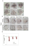
Most bilaterians exhibit a left-right asymmetric distribution of their internal organs. The sea urchin larva is notable in this regard since most adult structures are generated from left sided embryonic structures. The gene regulatory network governing this larval asymmetry is still a work in progress but involves several conserved signaling pathways including Nodal, and BMP. Here we provide a comprehensive analysis of Hedgehog signaling and it's contribution to left-right asymmetry. We report that Hh signaling plays a conserved role to regulate late asymmetric expression of Nodal and that this regulation occurs after Nodal breaks left-right symmetry in the mesoderm. Thus, while Hh functions to maintain late Nodal expression, the molecular asymmetry of the future coelomic pouches is locked in. Furthermore we report that cilia play a role only insofar as to transduce Hh signaling and do not have an independent effect on the asymmetry of the mesoderm. From this, we are able to construct a more complete regulatory network governing the establishment of left-right asymmetry in the sea urchin.
Keywords: Cilia; Hedgehog; Nodal; Right–left asymmetry; Sea urchin.
Copyright © 2016 Elsevier Inc. All rights reserved.
Figures


Similar articles
-
A conserved role for the nodal signaling pathway in the establishment of dorso-ventral and left-right axes in deuterostomes.J Exp Zool B Mol Dev Evol. 2008 Jan 15;310(1):41-53. doi: 10.1002/jez.b.21121. J Exp Zool B Mol Dev Evol. 2008. PMID: 16838294 Review.
-
Left-right asymmetry in the sea urchin embryo is regulated by nodal signaling on the right side.Dev Cell. 2005 Jul;9(1):147-58. doi: 10.1016/j.devcel.2005.05.008. Dev Cell. 2005. PMID: 15992548
-
Opposing nodal and BMP signals regulate left-right asymmetry in the sea urchin larva.PLoS Biol. 2012;10(10):e1001402. doi: 10.1371/journal.pbio.1001402. Epub 2012 Oct 9. PLoS Biol. 2012. PMID: 23055827 Free PMC article.
-
Telling left from right: left-right asymmetric controls in sea urchins.Genesis. 2014 Mar;52(3):269-78. doi: 10.1002/dvg.22739. Epub 2014 Jan 15. Genesis. 2014. PMID: 24395739 Review.
-
Hedgehog signaling patterns mesoderm in the sea urchin.Dev Biol. 2009 Jul 1;331(1):26-37. doi: 10.1016/j.ydbio.2009.04.018. Epub 2009 Apr 23. Dev Biol. 2009. PMID: 19393640 Free PMC article.
Cited by
-
The Power of Simplicity: Sea Urchin Embryos as in Vivo Developmental Models for Studying Complex Cell-to-cell Signaling Network Interactions.J Vis Exp. 2017 Feb 16;(120):55113. doi: 10.3791/55113. J Vis Exp. 2017. PMID: 28287557 Free PMC article.
-
Sea Urchin as a Universal Model for Studies of Gene Networks.Front Genet. 2021 Jan 20;11:627259. doi: 10.3389/fgene.2020.627259. eCollection 2020. Front Genet. 2021. PMID: 33552139 Free PMC article. Review.
-
Semi-automated, high-content imaging of drug transporter knockout sea urchin (Lytechinus pictus) embryos.J Exp Zool B Mol Dev Evol. 2024 May;342(3):313-329. doi: 10.1002/jez.b.23231. Epub 2023 Dec 12. J Exp Zool B Mol Dev Evol. 2024. PMID: 38087422 Free PMC article.
-
Intraflagellar transport: mechanisms of motor action, cooperation, and cargo delivery.FEBS J. 2017 Sep;284(18):2905-2931. doi: 10.1111/febs.14068. Epub 2017 Apr 18. FEBS J. 2017. PMID: 28342295 Free PMC article. Review.
-
Cilia are required for asymmetric nodal induction in the sea urchin embryo.BMC Dev Biol. 2016 Aug 23;16(1):28. doi: 10.1186/s12861-016-0128-7. BMC Dev Biol. 2016. PMID: 27553781 Free PMC article.
References
-
- Cameron RA, Fraser SE, Britten RJ, Davidson EH. The oral-aboral axis of a sea urchin embryo is specified by first cleavage. Development. 1989;106:641–647. - PubMed
Publication types
MeSH terms
Substances
Grants and funding
LinkOut - more resources
Full Text Sources
Other Literature Sources
Miscellaneous

