

Molecular basis of ion permeability in a voltage-gated sodium channel
- PMID: 26873592
- PMCID: PMC4972137
- DOI: 10.15252/embj.201593285
Molecular basis of ion permeability in a voltage-gated sodium channel
Abstract
Voltage-gated sodium channels are essential for electrical signalling across cell membranes. They exhibit strong selectivities for sodium ions over other cations, enabling the finely tuned cascade of events associated with action potentials. This paper describes the ion permeability characteristics and the crystal structure of a prokaryotic sodium channel, showing for the first time the detailed locations of sodium ions in the selectivity filter of a sodium channel. Electrostatic calculations based on the structure are consistent with the relative cation permeability ratios (Na(+) ≈ Li(+) ≫ K(+), Ca(2+), Mg(2+)) measured for these channels. In an E178D selectivity filter mutant constructed to have altered ion selectivities, the sodium ion binding site nearest the extracellular side is missing. Unlike potassium ions in potassium channels, the sodium ions in these channels appear to be hydrated and are associated with side chains of the selectivity filter residues, rather than polypeptide backbones.
Keywords: crystal structure; electrophysiology; ion permeability; sodium channel.
© 2016 The Authors. Published under the terms of the CC BY 4.0 license.
Figures
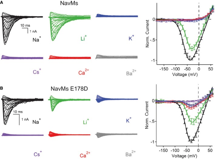
Left: Representative current traces from wild‐type NavMs channels showing the first 0.1 s of 0.5 s activations from −180 mV holding potential recorded with the indicated extracellular cations. Right: Resulting current–voltage profiles, where current magnitudes were normalised to the maximum inward current measured in the sodium condition (error ± SEM; n = 6–9).
Left: Representative current traces from mutant channel E178D (as in A). Right: Resulting current–voltage profiles (error ± SEM; n = 4–8).
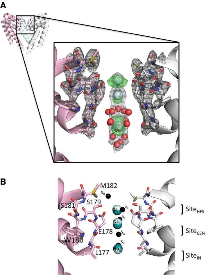
Upper: Overview of the AB tetramer, with each of the monomers depicted in a different colour. The front monomer has been removed for clarity. The black box indicates the region expanded in the lower panel. Lower: An expanded view of the selectivity filter, showing only two monomers as pink and white ribbon cartoons with the selectivity filter residues drawn as sticks in the corresponding colours. Sodium ions are shown as cyan spheres and well‐ordered waters as red spheres (at 0.4× their actual radii, for clarity). The final refined 2Fo‐Fc electron density maps for the protein, ions and water are shown at 2.0 σ in dark grey mesh and 0.8 σ in light grey, and the initial difference map (without sodium or waters included in the phase calculation) at 3.0 σ in green mesh.
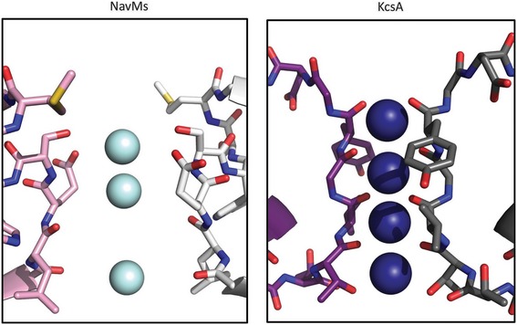
Superposition of the sodium ion binding sites found in the crystallographically distinct AB and CD tetramers and the proposed binding sites from other studies. The tetramers were superposed with the LSQKAB program (Kabsch, 2010) from CCP4 (Winn et al, 2011) using residues 131–220 from all four monomers in each tetramer. Only two monomers from the AB tetramer are shown for clarity, and coloured as in (A). Sodium ions from the AB tetramer are depicted as cyan spheres, those from the CD tetramer as teal spheres. Predicted sodium ion binding sites from Ulmschneider et al (2013) are indicated as small black spheres and labelled on the right of the spheres according to the nomenclature (S0–S4) in that study. Predicted sodium ion binding sites from Payandeh et al (2011), based on a closed channel structure of the close orthologue NavAb, are indicated on the far right and labelled as per the nomenclature of that study (SiteHFS, SiteCEN and SiteIN). This and subsequent molecular figures were drawn with PyMol (The PyMOL Molecular Graphics System, Version 1.6, Schrödinger, LLC) unless otherwise stated.
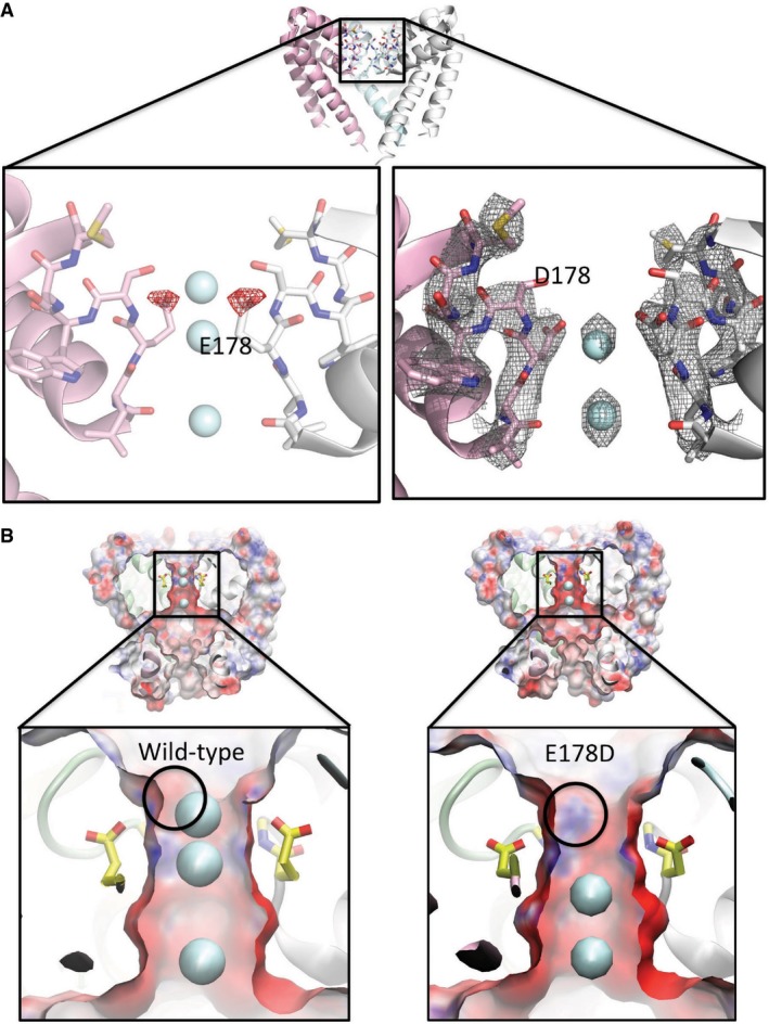
Top: Crystal structure overview of the wild‐type AB tetramer (PDBID 5BZB), view as in Fig 2A. Left: Difference (Fo–Fc) electron density map (mutant minus wild‐type) contoured at −2.5 σ and drawn as red mesh, plotted on the structure of the wild‐type (E178) protein, highlighting the consequences of the loss of the δ‐carbon atom from residue 178 in the E178D mutant. For reference, the locations of the sodium ion sites present in the wild‐type (E178) structure are depicted as cyan spheres. Both the structure and the ions are displayed as faded backgrounds so that the difference map can be seen more clearly. Right: Final refined 2Fo–Fc electron density map for the E178D mutant (PDBID 4X88), contoured at 1.5 σ and shown as grey mesh, with the sodium ions depicted as cyan spheres.
Surface representations of (left) wild‐type NavMs pore and (right) the E178D mutant coloured by electrostatic charge calculated by APBSmem (Callenberg et al, 2010) and drawn with VMD (Humphrey et al, 1996), showing a slice through the middle of the tetramer. Sodium ions are shown as cyan spheres; only 2 are present in the mutant, instead of the 3 seen in the wild‐type, with the top sodium missing in the mutant. The lower panels are expanded views of the selectivity filter regions, with the E178 side chains depicted in yellow sticks and the location of the main chain nitrogen of residue 179 indicated by the black circle. The alteration in the protein structure caused by removal of the methylene group of E178 exposes a positive region of the polypeptide backbone adjacent to the site where the top sodium ion is located in the wild‐type protein; this likely contributes to why an equivalent sodium ion is not seen adjacent to this position in the mutant.
Comment in
-
Three in a row-how sodium ions cross the channel.EMBO J. 2016 Apr 15;35(8):793-5. doi: 10.15252/embj.201694094. Epub 2016 Mar 21. EMBO J. 2016. PMID: 27002160 Free PMC article.
References
Publication types
MeSH terms
Substances
Grants and funding
- BB/L006790/BB_/Biotechnology and Biological Sciences Research Council/United Kingdom
- BB/L02625/BB_/Biotechnology and Biological Sciences Research Council/United Kingdom
- T32 HL007572/HL/NHLBI NIH HHS/United States
- T32-HL007572/HL/NHLBI NIH HHS/United States
- K99 DK106655/DK/NIDDK NIH HHS/United States
LinkOut - more resources
Full Text Sources
Other Literature Sources
Miscellaneous
