Review
. 2016 Sep;73(9):477-97.
doi: 10.1002/cm.21282. Epub 2016 Mar 1.

Actin dynamics and cofilin-actin rods in alzheimer disease

Affiliations
Review

Actin dynamics and cofilin-actin rods in alzheimer disease

James R Bamburg et al. Cytoskeleton (Hoboken). 2016 Sep.

Abstract

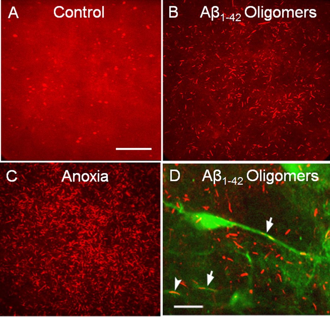
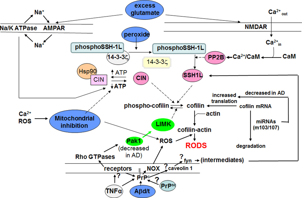
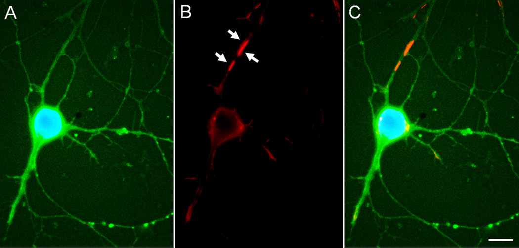
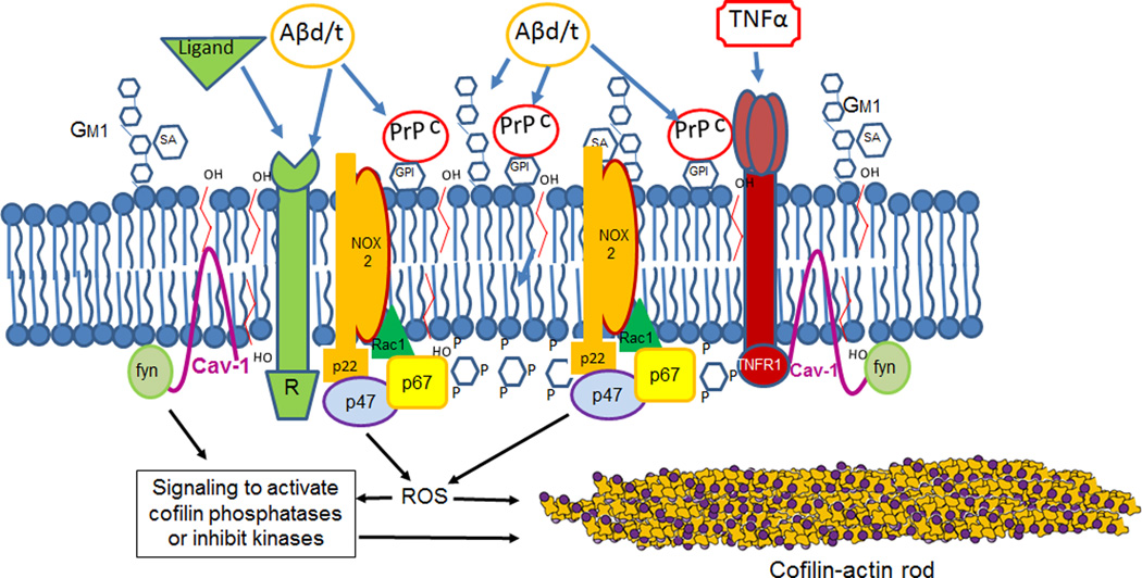
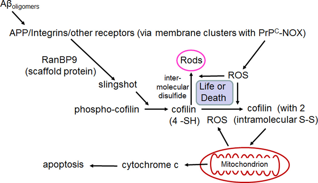
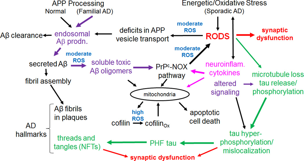
Cytoskeletal abnormalities and synaptic loss, typical of both familial and sporadic Alzheimer disease (AD), are induced by diverse stresses such as neuroinflammation, oxidative stress, and energetic stress, each of which may be initiated or enhanced by proinflammatory cytokines or amyloid-β (Aβ) peptides. Extracellular Aβ-containing plaques and intracellular phospho-tau-containing neurofibrillary tangles are postmortem pathologies required to confirm AD and have been the focus of most studies. However, AD brain, but not normal brain, also have increased levels of cytoplasmic rod-shaped bundles of filaments composed of ADF/cofilin-actin in a 1:1 complex (rods). Cofilin, the major ADF/cofilin isoform in mammalian neurons, severs actin filaments at low cofilin/actin ratios and stabilizes filaments at high cofilin/actin ratios. It binds cooperatively to ADP-actin subunits in F-actin. Cofilin is activated by dephosphorylation and may be oxidized in stressed neurons to form disulfide-linked dimers, required for bundling cofilin-actin filaments into stable rods. Rods form within neurites causing synaptic dysfunction by sequestering cofilin, disrupting normal actin dynamics, blocking transport, and exacerbating mitochondrial membrane potential loss. Aβ and proinflammatory cytokines induce rods through a cellular prion protein-dependent activation of NADPH oxidase and production of reactive oxygen species. Here we review recent advances in our understanding of cofilin biochemistry, rod formation, and the development of cognitive deficits. We will then discuss rod formation as a molecular pathway for synapse loss that may be common between all three prominent current AD hypotheses, thus making rods an attractive therapeutic target. © 2016 Wiley Periodicals, Inc.

Keywords: NADPH oxidase; amyloid-β; oxidative stress; prion signaling; proinflammatory cytokines.


Conflict of interest statement

The authors have no conflict of interest to declare.

Figures

Figure 1
Figure 1. Cofilin-stained rods in cultured mouse hippocampal slices induced by treatment with Aβ peptides or anoxia
Projection images of confocal stacks (30 µm) of cofilin immunostained cultured mouse hippocampal brain slices (14 d in vivo before treatment). (a) Untreated slice showing diffuse cofilin labelling and some puntate spheroid staining. (b) Slice treated for 24 h with 600 nM Aβ1–42 oligomers. (c) Brain slice subjected to one hour of anoxia before fixation. (d) Brain slice from a Thy-1-YFP-transgenic mouse treated similarly to the slice in (b). Arrows point to YFP-positive neurites with rods. Only ~10% of neurons in the hippocampus of the Thy-1-YFP mice express YFP. For all panels, slices were fixed in 4% formaldehyde, permeabilized with 100% methanol (−20° C) for 3 min, blocked and immunostained for cofilin with rabbit 1439 antibody [Shaw et al., 2004] and an Alexa 561 or 594 secondary antibody. Scale bars (a–c) and (d) 10 µm. Images modified from Davis et al., 2009.
Figure 2
Figure 2. Schematic of likely molecular routes of rod formation from cofilin and actin pools
Several routes of rod formation are possible, including oxidative cross-linking of cofilin before or after binding to F-actin or to actin monomers to induce rod assembly. Profilin binding to actin monomers opens the nucleotide pocket allowing the actin-bound nucleotide to equilibrate with the cellular adenine nucleotide pool, whereas cofilin binding to ADP-actin inhibits nucleotide exchange [Nishida, 1985].
Figure 3
Figure 3. Schematic of abbreviated signaling pathways contributing to rod formation by some initiators of neuronal stress and factors that change during AD progression
The role of caveolin and fyn in rod formation is hypothetical but is based on their known PrPC-mediated phosphorylation [Shi et al., 2013] and that fyn inhibitors can rescue synapse loss and established memory [Kaufman et al., 2015]. Other steps diagrammed here are discussed in the text.
Figure 4
Figure 4. Transmission electron micrographs of a rod-containing neurite showing the ~9 nm filament components in a cultured hippocampal slice treated 24 hr with Aβ1–42 oligomers
Slices were fixed, sectioned (250 nm) and stained as described elsewhere [Davis et al., 2009]. A rod runs the entire length of the neurite in the image shown (from lower left to upper right). Filaments of the rod surround a mitochondrion. White arrow points to 12 nm gold particle (slightly wider than the filament on which it sits), which was used as a fiduciary marker in reconstructing a tilted image stack to give the tomogram in panel (b). Scale bar = 100 nm.
Figure 5
Figure 5. Overexpression of EGFP-PrPC induces rods in cultured neurons
Cultured hippocampal rat E18 neuron (6 days in vitro) infected on day 3 with 100 moi of adenovirus for expression of EGF-PrPC (green), fixed on day 6, and immunostained for cofilin (red) and stained for DNA with DAPI (blue). (a) EGFP and DAPI stained image of neuron. (b) Cofilin immunostained cell shown in (a). White arrows point to rods. (c) Overlay of (a) and (b). Scale bar =10 µm. Images courtesy of L.S. Minamide.
Figure 6
Figure 6. Schematic of a PrPC- and sphingolipid-enriched membrane signaling domain
A number of receptors are found in these domains, some of which have been demonstrated to require PrPC as a co-receptor for Aβ-mediated rod signaling. We propose that because overexpression of PrPC alone drives rod formation through a NOX-dependent step [Walsh et al., 2014], multiple Aβ-binding components of the signaling complexes work in conjunction with PrPC to elevate the neurite ROS levels above the threshold required for rod-induction and maintenance. Other components of the signaling complex such as fyn [Ochs and Malaga-Trillo, 2014], or perhaps ROS itself (see Figure 3), activate cofilin.
Figure 7
Figure 7. Schematic of pathway for Aβ oligomer-induced rods in neurons through the β1-integrin receptor and RanBP9 scaffolding protein
We hypothesize that the formation of intermolecular disulfide bonds in cofilin (leading to rod formation) in response to ROS production is competitive with cofilin's intramolecular oxidation, which targets it to mitochondria to trigger apoptosis. Rods are reversible if REDOX pathways can reduce the ROS levels and thus rods may form and disappear many times. We hypothesize that eventually a large surge in ROS may trigger cofilin-induced apoptosis occurring later in AD progression.
Figure 8
Figure 8. A hypothetical model in which rods play a central role in mediating AD progression
Intermediates or initiators for the three major hypotheses for AD are shown in color: purple for the amyloid hypothesis, green for the tauopathy hypothesis and fuscia for the neuroinflammatory/cytokine hypothesis. Sites where reactive oxygen species are thought to be directly involved are in blue. In cognitively normal individuals, Aβ produced in endosomes is cleared or it may form fibrils and be sequestered into extracellular plaques in the brain. Although found in familial and sporadic AD, plaques may also be found in cognitively normal subjects and thus by themselves do not cause dementia. Changes in the form of soluble Aβ, arising from oxidation or other modifications, produce a more rod-inducing and synaptotoxic form. Other initiators of sporadic AD, such as neuroinflammatory cytokines, initiate a moderate ROS response that leads to rods and greater amounts of Aβ production in a feed-forward pathway. We hypothesize that rod-induced microtubule disruption releases tau for phosphorylation and initiation of PHF-tau formation, although this could be directly mediated by altered signaling. Mitochondrial dysfunction resulting from many possible sources eventually produces high ROS levels that contribute to cofilin oxidation (two internal disulfide bonds) that target it to mitochondria and cause release of cytochrome c, initiating apoptosis. Since rods are reversible, cofilin-induced apoptosis may gradually increase during cycles of rod reversal and acute high ROS levels.

References

    1. Agnew BJ, Minamide LS, Bamburg JR. Reactivation of phosphorylated actin depolymerizing factor and identification of the regulatory site. J Biol Chem. 1995;270:17582–17587. - PubMed
    1. Agostini F, Dotti CG, Perez-Canamas A, Ledesma MD, Benetti F, Legname G. Prion protein accumulation in lipid rafts of mouse aging brain. PLOS ONE. 2013;8:e74244. - PMC - PubMed
    1. Alzheimer’s Association. 2015 Alzheimer’s disease facts and figures. Alzheimer’s & Dementia. 2015;11:332–384. - PubMed
    1. Andrianantoandro E, Pollard TD. Mechanism of actin filament turnover by severing and nucleation at different concentrations of ADF/cofilin. Mol Cell. 2006;24:13–23. - PubMed
    1. Andziak B, Buffenstein R. Disparate patterns of age-related changes in lipid peroxidation in long-lived naked mole-rats and shorter lived mice. Aging Cell. 2006;5:525–532. - PubMed

MeSH terms