

External fixation reconstruction of the residual problems of benign bone tumours
- PMID: 26873644
- PMCID: PMC4814386
- DOI: 10.1007/s11751-016-0244-8
External fixation reconstruction of the residual problems of benign bone tumours
Erratum in
-
Erratum to: External fixation reconstruction of the residual problems of benign bone tumours.Strategies Trauma Limb Reconstr. 2016 Apr;11(1):51. doi: 10.1007/s11751-016-0248-4. Strategies Trauma Limb Reconstr. 2016. PMID: 26993112 Free PMC article. No abstract available.
Abstract
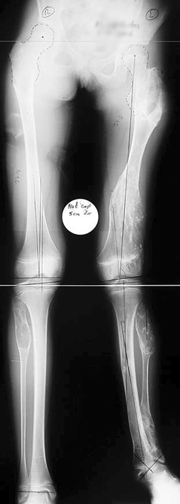
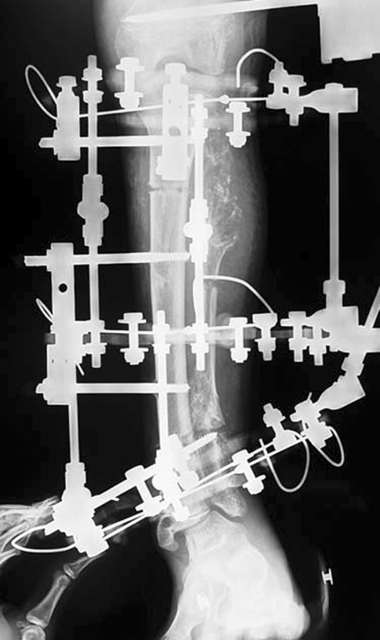
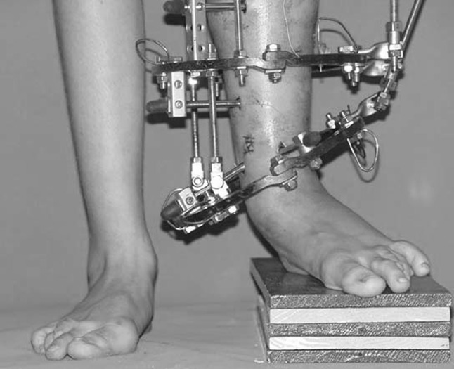
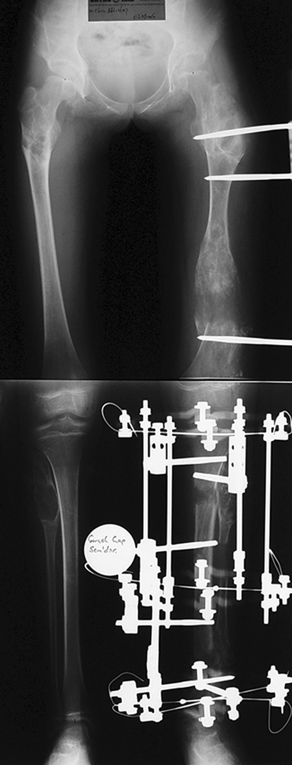
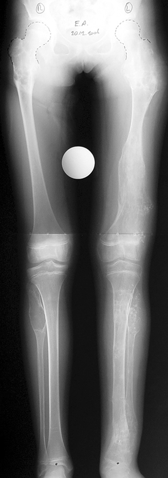
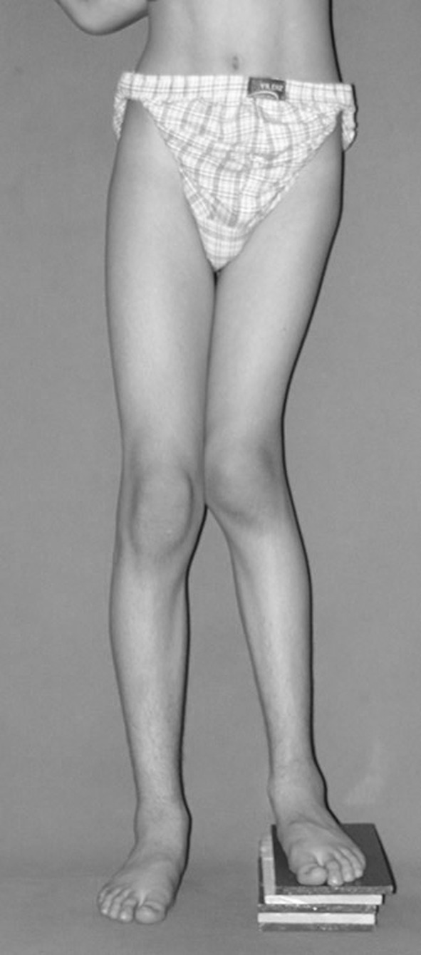
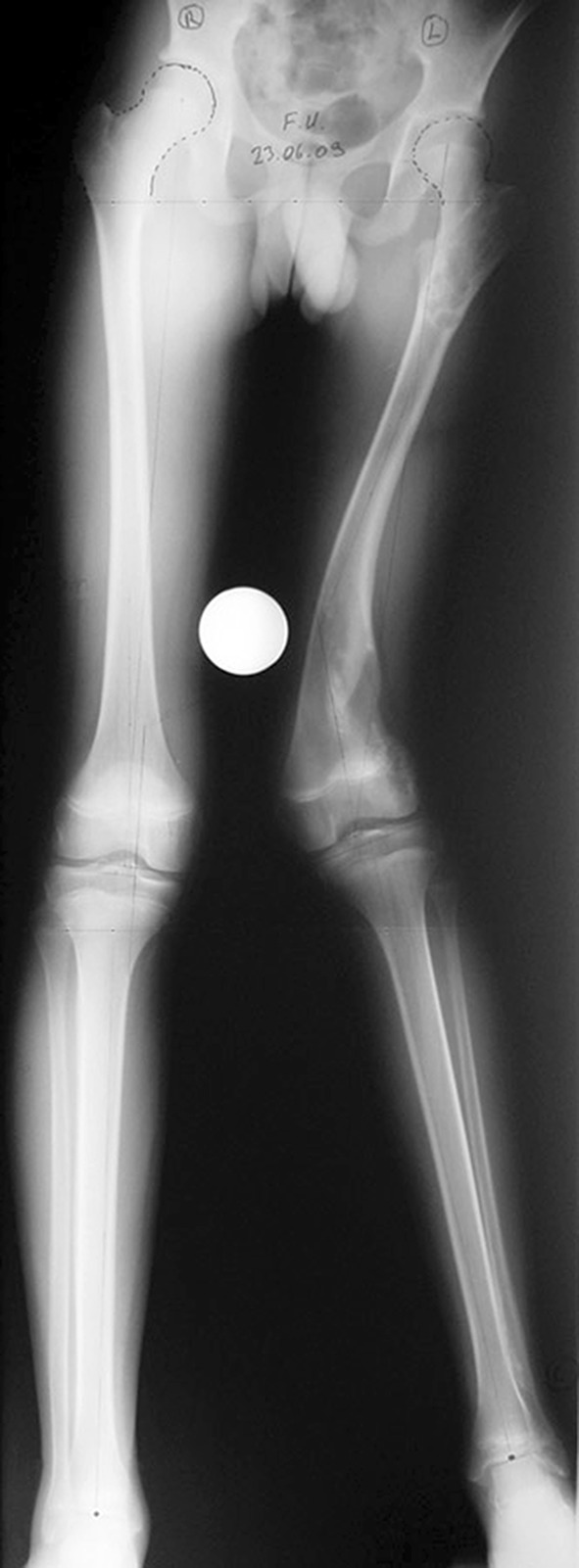
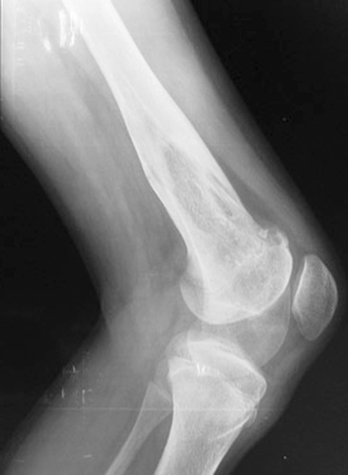
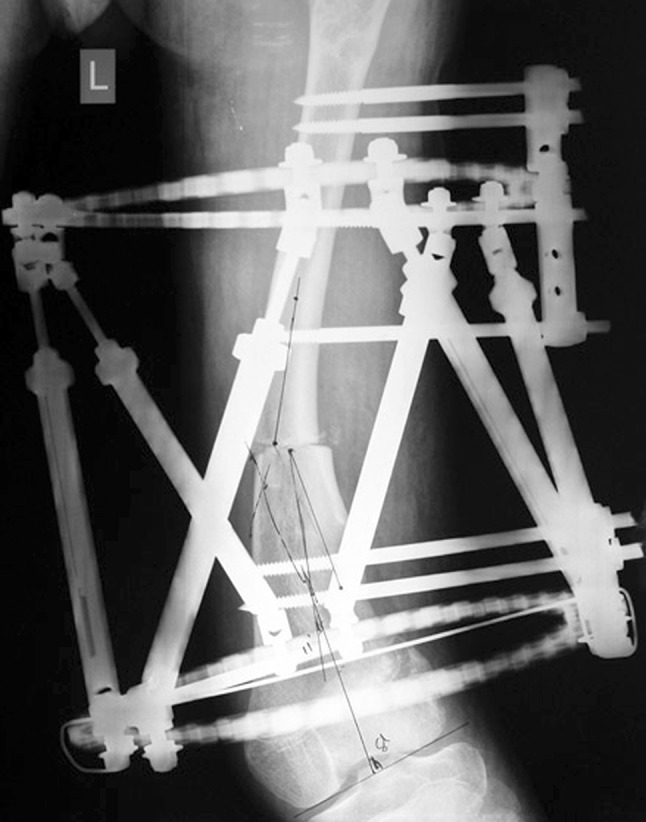
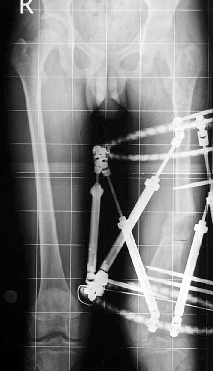
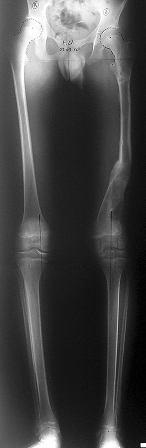
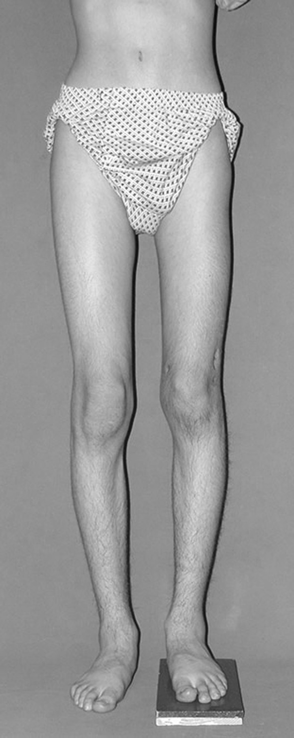
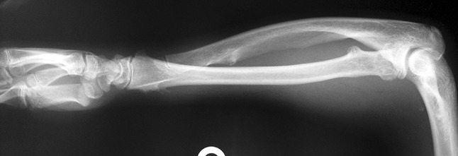
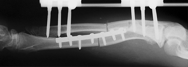
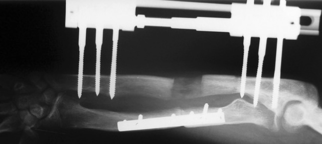
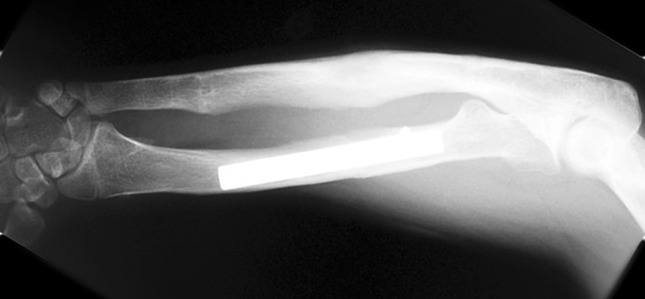
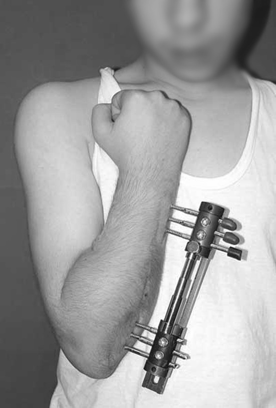
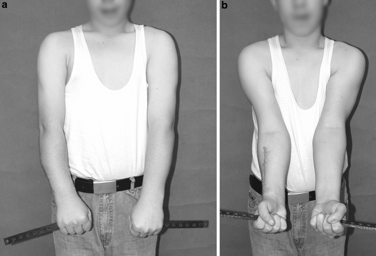
The mechanical features of and biologic response to using distraction osteogenesis with the circular external fixator are the unique aspects of Ilizarov's contribution that allows deformity correction and reconstruction of bone defects. We present a retrospective study of 20 patients who suffered from a variety of benign tumours for which external fixators (EF) were used to treat deformity, bone loss, and limb-length discrepancy. A total of 26 bony segments in twenty patients (10 males, 10 females; mean age 17 years; range 7-58 years) were treated with EF for residual problems from the tumour itself (primary treatment) in 8 patients and for complications related to the primary surgery (secondary treatment) in 12 patients. Histological diagnoses were Ollier's disease (n = 4), Fibrous Dysplasia (n = 5), Congenital multiple exostosis (n = 5), giant cell tumour (n = 2) and one case for chondromyxoid fibroma, desmoid fibroma, chondroma and unicameral bone cyst. Various types of external fixators used to treat these problems. These were Ilizarov, unilateral fixator, multiaxial correction frame (Biomet, Parsippany, NJ), Taylor spatial frame (Memphis, TN) and smart correction multiaxial frame. The mean follow-up time was 69.5 months (range 35-108 months). The mean external fixation time was 159.5 days (range 27-300 days). The mean external fixation index was 67.4 days/cm (12-610) in 26 limbs who underwent distraction osteogenesis. The mean length of distraction was 4.9 cm (range 0.2-14 cm). At final follow-up, all patients had returned to normal activities. Complications were in the form of knee arthrodesis in one patient, pin tract infection in six and residual shortening in eight patients. The use of EF and the principles of distraction osteogenesis, in the management of problems associated with benign bone tumours and related surgery yields successful results especially in young patients. With this approach, the risk for recurrence of shortening and deformity may be minimized with overcorrection or over-lengthening as dictated by preoperative planning.
Keywords: Benign bone tumours; Bone deformity; Distraction osteogenesis; External fixation; Limb reconstruction; Shortening.
Figures
Similar articles
-
[Applications of external fixation for management of complications associated with musculoskeletal tumors and related surgery].Acta Orthop Traumatol Turc. 2009 May-Jul;43(3):219-28. doi: 10.3944/AOTT.2009.219. Acta Orthop Traumatol Turc. 2009. PMID: 19717939 Turkish.
-
Treatment of benign bone tumours using external fixation.J Bone Joint Surg Br. 2007 Aug;89(8):1077-83. doi: 10.1302/0301-620X.89B8.19132. J Bone Joint Surg Br. 2007. PMID: 17785749
-
Bifocal compression-distraction in the acute treatment of grade III open tibia fractures with bone and soft-tissue loss: a report of 24 cases.J Orthop Trauma. 2004 Mar;18(3):150-7. doi: 10.1097/00005131-200403000-00005. J Orthop Trauma. 2004. PMID: 15091269
-
Limb lengthening for deformities in Ollier's disease: a systematic review.Eur J Orthop Surg Traumatol. 2020 Dec;30(8):1325-1332. doi: 10.1007/s00590-020-02692-5. Epub 2020 Jun 4. Eur J Orthop Surg Traumatol. 2020. PMID: 32500348
-
External fixators: looking beyond the hardware maze.Skeletal Radiol. 2020 Mar;49(3):359-374. doi: 10.1007/s00256-019-03306-w. Epub 2019 Sep 12. Skeletal Radiol. 2020. PMID: 31515594 Review.
Cited by
-
Role of the Ilizarov non-free bone plasty in the management of long bone defects and nonunion: Problems solved and unsolved.World J Orthop. 2020 Jun 18;11(6):304-318. doi: 10.5312/wjo.v11.i6.304. eCollection 2020 Jun 18. World J Orthop. 2020. PMID: 32572367 Free PMC article.
-
Current paediatric orthopaedic practice in hereditary multiple osteochondromas of the forearm: a systematic review.SICOT J. 2018;4:10. doi: 10.1051/sicotj/2018002. Epub 2018 Mar 21. SICOT J. 2018. PMID: 29565244 Free PMC article.
-
Limb Length Discrepancy and Angular Deformity due to Benign Bone Tumors and Tumor-like Lesions.J Am Acad Orthop Surg Glob Res Rev. 2021 Mar 10;5(3):e00214. doi: 10.5435/JAAOSGlobal-D-20-00214. J Am Acad Orthop Surg Glob Res Rev. 2021. PMID: 33720060 Free PMC article.
-
Lower limb lengthening and deformity correction in polyostotic fibrous dysplasia using external fixation and flexible intramedullary nailing.J Orthop. 2020 Mar 28;21:192-198. doi: 10.1016/j.jor.2020.03.014. eCollection 2020 Sep-Oct. J Orthop. 2020. PMID: 32256003 Free PMC article.
-
New bone formation accelerates during lower limb lengthening and deformity correction in children with Ollier's disease.J Orthop Traumatol. 2023 Jul 31;24(1):39. doi: 10.1186/s10195-023-00717-3. J Orthop Traumatol. 2023. PMID: 37524995 Free PMC article.
References
-
- Tsuchiya H, Tomita K, Shinokawa Y, et al. The Ilizarov method in the management of giant-cell tumors of the proximal tibia. J Bone Joint Surg [Br] 1996;78:264–269. - PubMed
-
- Tsuchiya H, Tomita K, Minematsu K, et al. Limb salvage using distraction osteogenesis: a classification of the technique. J Bone Joint Surg [Br] 1996;78:403–411. - PubMed
LinkOut - more resources
Full Text Sources
Other Literature Sources
