. 2017 Feb;16(1):203-229.
doi: 10.1007/s12311-016-0763-3.

Consensus Paper: Towards a Systems-Level View of Cerebellar Function: the Interplay Between Cerebellum, Basal Ganglia, and Cortex

Affiliations

Consensus Paper: Towards a Systems-Level View of Cerebellar Function: the Interplay Between Cerebellum, Basal Ganglia, and Cortex

Daniele Caligiore et al. Cerebellum. 2017 Feb.

Abstract

Despite increasing evidence suggesting the cerebellum works in concert with the cortex and basal ganglia, the nature of the reciprocal interactions between these three brain regions remains unclear. This consensus paper gathers diverse recent views on a variety of important roles played by the cerebellum within the cerebello-basal ganglia-thalamo-cortical system across a range of motor and cognitive functions. The paper includes theoretical and empirical contributions, which cover the following topics: recent evidence supporting the dynamical interplay between cerebellum, basal ganglia, and cortical areas in humans and other animals; theoretical neuroscience perspectives and empirical evidence on the reciprocal influences between cerebellum, basal ganglia, and cortex in learning and control processes; and data suggesting possible roles of the cerebellum in basal ganglia movement disorders. Although starting from different backgrounds and dealing with different topics, all the contributors agree that viewing the cerebellum, basal ganglia, and cortex as an integrated system enables us to understand the function of these areas in radically different ways. In addition, there is unanimous consensus between the authors that future experimental and computational work is needed to understand the function of cerebellar-basal ganglia circuitry in both motor and non-motor functions. The paper reports the most advanced perspectives on the role of the cerebellum within the cerebello-basal ganglia-thalamo-cortical system and illustrates other elements of consensus as well as disagreements and open questions in the field.

Keywords: Basal ganglia cerebellum anatomical link; Cerebellar motor and cognitive function; Movement disorders; Non-invasive brain stimulation; Nucleo-olivary inhibition; Parkinson’s disease tremor.


Figures
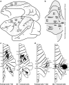
Fig. 1
Fig. 1
Cerebellar networks with the cerebral cortex. a The relative density of cerebral cortex neurons that project to the pontine nuclei is indicated by gray dots on the lateral and medial views of the monkey brain (based on [48]). Black labels indicate areas of the cerebral cortex that are the target of cerebellar output. Gray labels indicate areas that are not targets of cerebellar output (adapted from [3]). b Summary map of dentate nucleus output topography. The lettering on the unfolded map indicates the neocortical target of different output channels. The location of different output channels divides the dentate nucleus into motor and non-motor domains, separated by the dotted line (adapted from [45]). c Organization of cerebellar circuits with M1. Left: the distribution of Purkinje cells (small dots) that project to the arm area of M1. Right: the distribution of granule cells (fine lines) that receive input from the arm area of M1 (adapted from [50]). d Organization of cerebellar loops with area 46. Left: the distribution of Purkinje cells (small dots) that project to area 46. Right: the distribution of granule cells (fine lines) that receive input from area 46 (adapted from [50]). Numbers refer to cytoarchitectonic areas (a, b). Roman numerals refer to cerebellar lobules (c, d). Labels in italics refer to cortical sulci (a) and cerebellar fissures (c, d). AIP anterior intraparietal area, AS arcuate sulcus, CgS cingulate sulcus, FEF frontal eye field, IP intraparietal sulcus, LS lateral sulcus, Lu lunate sulcus, M1 face, arm, and leg areas of the primary motor cortex, PMd arm arm area of the dorsal premotor area, PMv arm arm area of the ventral premotor area, PrePMd predorsal premotor area, PreSMA presupplementary motor area, PS principal sulcus, SMA arm arm area of the supplementary motor area, ST superior temporal sulcus, TE area of inferotemporal cortex
Fig. 2
Fig. 2
Cerebellar interconnections with the basal ganglia. Based on [12] and [13]. DN dentate nucleus, GPe external segment of the globus pallidus, GPi internal segment of the globus pallidus, PN pontine nuclei, STN subthalamic nucleus
Fig. 3
Fig. 3
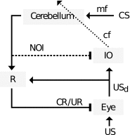
Cerebellar circuitry of the eyeblink response, with NOI. An air puff (US) is detected at the eye and elicits a neural response (USd) that by recruiting the reflex (R, oculomotor neurons) triggers the feedback reaction (UR). The effect of USd decreases proportionally to the degree of eyelid closure. The internally generated USd signal first reaches the IO and, subsequently, the cerebellum via the climbing fibers (cf). The convergence of the US signal with the CS information entering the cerebellum through the mossy fiber (mf) pathway induces plasticity in the cerebellar cortex, causing the inhibitory Purkinje cells to gradually develop a long-latency pause in their simple spikes firing. This acquired pause disinhibits target neurons in the cerebellar deep nuclei which provide the output of the circuit. This output not only reaches the downstream reflex controller (R), triggering an anticipatory/feed-forward response (CR), but also, via the NOI, counteracts the activation of the IO by the USd. In consequence, and assuming that learning stabilizes once IO activity remains at baseline, the USd signal should not be completely abolished peripherally; otherwise, the NOI would depress the IO firing below baseline introducing a negative error, which would lead to the extinction of the CR. Note that the excitatory and inhibitory outputs of the cerebellum are generated by distinct neuronal populations. Triangular arrowheads indicate excitatory effects and rectangular inhibitory ones. The CR(s)/UR(s) reaching the eye motor plant are depicted as inhibitory as they diminish the sensory consequences of the air puff US. The dotted line indicates the error signal, and the dashed line indicates a delayed connection. Note that even though we have only indicated the CS signal in the mossy fiber pathway, rich multisensory information reaches the cerebellar cortex through that pathway including, e.g., the US signal. However, by definition, only the CS will be of use in order to predict the US
Fig. 4
Fig. 4
The DPM architecture for learning and control. Functionally related areas of the cerebral cortex communicate with each other via corticocortical loops [104]. In addition, most cortical areas have important learning and control loops with subcortical structures, particularly with the basal ganglia (BG) and with the cerebellum (CB). Evidence for relatively private loops through BG and through CB comes from many neuroscientific studies (e.g., [, –98]). The rectangle outlines one distributed processing module or DPM [96]. It includes one area of cerebral cortex together with its loops of connectivity through BG and CB. The different DPMs communicate with each other mainly through reciprocal corticocortical loops
Fig. 5
Fig. 5
Learning and control operations in a DPM. Hebbian learning occurs in cerebral cortex. The control operation is pattern formation. Reinforcement learning occurs in basal ganglia (BG). The main control operation is pattern classification, which occurs in the striatum on cortical and thalamic input to spiny projection neurons (SPNs). Through direct (disinhibition) and indirect (inhibition of disinhibition) pathways, a coarse selection of goals is briefly stored in reciprocal corticothalamic pathways. Supervised learning occurs in the refinement stage in the cerebellar cortex, through depression of parallel fiber/Purkinje cell synapses. The positive feedback loop between the cerebellar nucleus and cerebral cortex is a working memory that is regulated by potent inhibition from Purkinje cells
Fig. 6
Fig. 6
A possible implementation of model-based action selection by combining forward models in the cerebellum and reward predictors in the basal ganglia. From [111] with permission
Fig. 7
Fig. 7
Functional connectivity within a network encompassing cerebellar nuclei, the thalamus, and basal ganglia was significantly greater than at baseline 30 min after exposure to an implicit sequence learning task. Similar connectivity increase was seen for explicit learning; in both cases, this declined after 6 h. From [155] with permission
Fig. 8
Fig. 8
Schematic representation of the known main communication pathways among the equally important three nodes of the motor control network
Fig. 9
Fig. 9
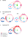
Schematic representation of the basal ganglia-thalamo-cortical loop, the cerebello-thalamo-cortical loop, and the interaction between the two in health (a), in non-dyskinetic Parkinson’s disease, after levodopa withdrawal (OFF) and after regular dose of levodopa (ON) (b), and in advanced Parkinson’s disease with levodopa-induced dyskinesia (c). Red arrows represent glutamatergic projections; blue arrows represent GABA-ergic projections; green arrows represent dopaminergic projections; dark green arrows in panels b and c represent the exogenous dopamine from levodopa. The shades of the blocks represent the activity of the respective network nodes. The STN is overactive because of cortical glutamatergic over activity during dyskinesias and from loss of GPe inhibition in OFF. The STN over activity locks cerebellar cortex in a persistent hyperactive state and interferes with its sensory processing function. The behavior of the cortico-ponto-cerebellar projections in non-dyskinetic PD in ON is not reported so far and is predicted by this model to be close to normal (CB ctx cerebellar cortex, CM centromedian thalamic nucleus, D1/D2 dopamine receptor types of the striatal medium spiny neurons, DN dentate nucleus, GPe globus pallidus externus, GPi globus pallidus internus, M1 primary motor cortex, PF parafascicular nucleus, PMC premotor cortex, PN pontine nuclei, SMA supplementary motor area, SNc substantia pars compacta, SNr substantia pars reticulata, STN subthalamic nucleus, VL ventrolateral thalamic nucleus, VLPo ventro-latero-posterior thalamic nucleus pars oralis, VTA ventral tegmental area)
Fig. 10
Fig. 10
Figure showing the basal ganglia (in black) and the cerebello-thalamic pathway (in light gray) including the motor cortex (in dark gray) where both circuits anatomically converge. Both the basal ganglia and the cerebello-thalamic loop are involved in the production of Parkinson’s resting tremor, but it is unclear how they interact. Here we show four possible anatomical pathways through which the basal ganglia may influence the cerebello-thalamic circuit (colored lines). Pathway 1 connects both circuits through the pons (blue), pathway 2 through the motor cortex (red), pathway 3 through the zona incerta (green), and pathway 4 through the peripheral muscles (orange). CBLM cerebellum, GPi globus pallidus internus, GPe globus pallidus externus, ILN interlaminar nuclei, MC motor cortex, STN subthalamic nucleus, VLa anterior part of venterolateral nucleus of thalamus, VLp posterior part of venterolateral nucleus of thalamus, VIM ventral intermediate nucleus of thalamus, ZI zona incerta

Comment in

References

    1. Strick PL, Dum RP, Fiez JA. Cerebellum and nonmotor function. Annu Rev Neurosci. 2009;32:413–434. doi: 10.1146/annurev.neuro.31.060407.125606. - DOI - PubMed
    1. Koziol LF, Budding DE, Chidekel D. From movement to thought: executive function, embodied cognition, and the cerebellum. Cerebellum. 2012;11(2):505–525. doi: 10.1007/s12311-011-0321-y. - DOI - PubMed
    1. Bostan AC, Dum RP, Strick PL. Cerebellar networks with the cerebral cortex and basal ganglia. Trends Cogn Sci. 2013;17(5):241–254. doi: 10.1016/j.tics.2013.03.003. - DOI - PMC - PubMed
    1. Caligiore D, Pezzulo G, Miall RC, Baldassarre G. The contribution of brain sub-cortical loops in the expression and acquisition of action understanding abilities. Neurosci Biobehav R. 2013;37(10):2504–2515. doi: 10.1016/j.neubiorev.2013.07.016. - DOI - PMC - PubMed
    1. Alexander GE, DeLong MR, Strick PL. Parallel organization of functionally segregated circuits linking basal ganglia and cortex. Annu Rev Neurosci. 1986;9(1):357–381. doi: 10.1146/annurev.ne.09.030186.002041. - DOI - PubMed

Publication types