. 2016 Apr 20;44(7):3390-407.
doi: 10.1093/nar/gkw074. Epub 2016 Feb 11.

Initiation on the divergent Type I cadicivirus IRES: factor requirements and interactions with the translation apparatus

Affiliations

Initiation on the divergent Type I cadicivirus IRES: factor requirements and interactions with the translation apparatus

Mukta Asnani et al. Nucleic Acids Res. .

Abstract

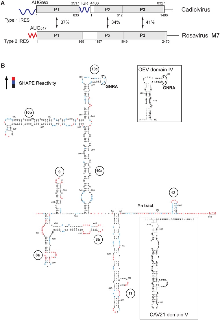
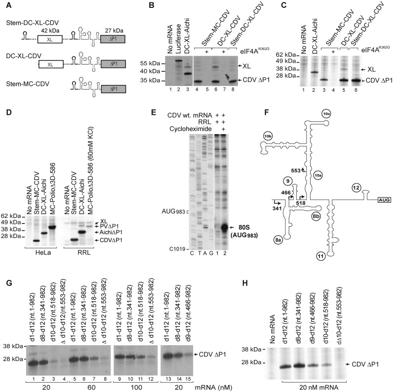
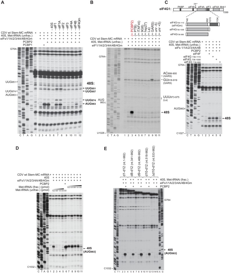
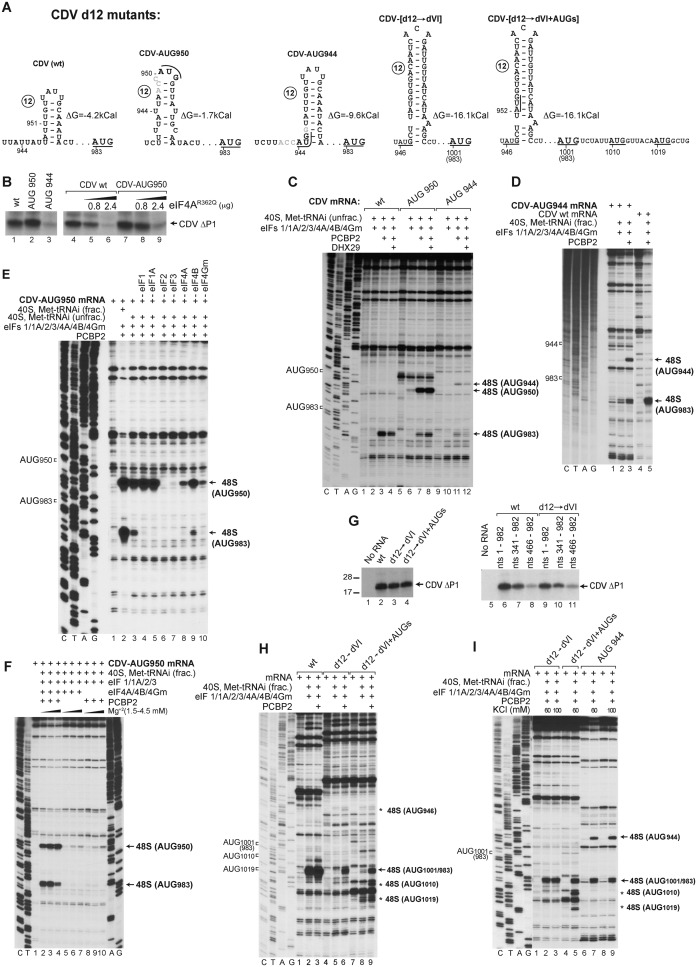
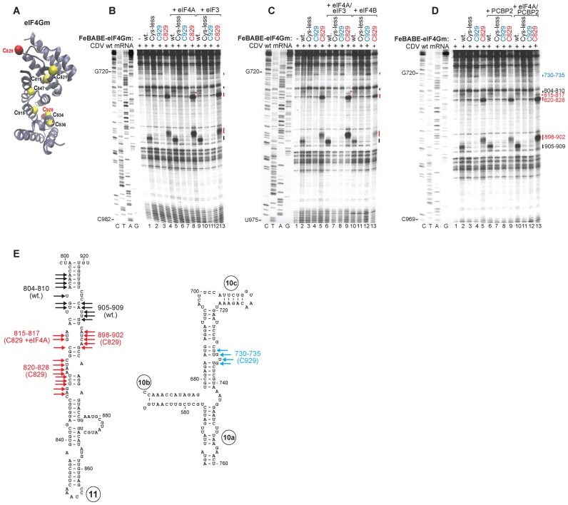
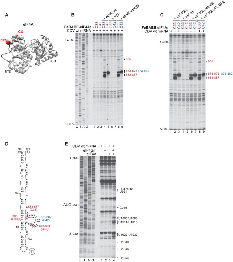
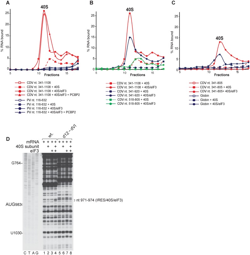
Cadicivirus (CDV) is unique amongst picornaviruses in having a dicistronic genome with internal ribosomal entry sites (IRESs) preceding both open reading frames. Here, we investigated initiation on the 5'-terminal IRES. We report that the 982-nt long 5'UTR comprises 12 domains (d1-d12), five of which (d8-d12, nts 341-950) constitute a divergent Type I IRES. It comprises central elements (the apex of d10, d11 and the following polypyrimidine tract) that are homologous to corresponding elements in canonical Type 1 IRESs, and non-canonical flanking domains (d8, d9 and d12). In vitro reconstitution revealed that as with canonical Type I IRESs, 48S complex formation requires eukaryotic initiation factors (eIFs) 1, 1A, 2, 3, 4A, 4B and 4G, and the poly(C) binding protein 2 (PCBP2), and starts with specific binding of eIF4G/eIF4A to d11. However, in contrast to canonical Type I IRESs, subsequent recruitment of 43S ribosomal complexes does not require direct interaction of their eIF3 constituent with the IRES-bound eIF4G. On the other hand, the CDV IRES forms a 40S/eIF3/IRES ternary complex, with multiple points of contact. These additional interactions with translational components could potentially stimulate recruitment of the 43S complex and alleviate the necessity for direct eIF4G/eIF3 interaction.


Figures

Figure 1.
Figure 1.
Structure and genomic context of the CDV 5’UTR IRES. (A) Schematic representations of CDV strain 209 and Rosavirus M7 genomes, showing the 5’UTR (including the IRES), P1 structural and P2-P3 non-structural protein coding regions, the 3’UTR and in CDV, the intergenic region (IGR), and labeled to indicate amino acid sequence identity between homologous regions. Numbers shown above and below the borders of domains refer to nucleotides and amino acid residues, respectively. (B) Secondary structure model of the CDV strain 209 IRES, color-coded to show reactivity to NMIA (see key at upper left and data shown in Supplementary Figure S1). The initiation codon (AUG983) is underlined. The upper inset panel shows the apical region of dIV of the Type I Ovine enterovirus IRES, showing the GNRA tetraloop and sequence identity with the corresponding element of CDV subdomain 10c (bold font). The lower inset panel shows the apical region of dV and the Yn tract of the Type I IRES of Coxsackievirus A21, showing sequence identity with CDV d11 and downstream sequences (bold font).
Figure 2.
Figure 2.
The 5’ border of the CDV 5’UTR IRES. (A) Schematic representation of monocistronic (MC) and dicistronic (DC) CDV mRNAs, with the CDV 5’UTR and a stable hairpin (thick line) placed upstream of the first and/or the second cistron. (B-D) Translational activity of the IRES in (B and D) RRL and (C and D) HeLa cell-free translation extract, and its sensitivity to inhibition by dominant-negative eIF4AR362Q. Luciferase and DC Aichivirus mRNAs (31) were used as controls for the efficiency of translation. (E) Toe-print analysis of 80S ribosomes formed on wt CDV MC mRNA in RRL in the presence of cycloheximide. (F) Model of the IRES, with arrows indicating the borders of 5’-terminal truncations of the 5’UTR. (G and H) Effect of these truncations, introduced into Stem-MC mRNA, on IRES activity in (G) RRL and (H) HeLa cell-free translation extract.
Figure 3.
Figure 3.
Factor requirements for initiation on the CDV IRES. Toe-printing analysis of 48S complex formation on CDV MC mRNAs containing (AD) full-length or (E) 5’-terminally truncated derivatives of the 5’UTR, in the in vitro reconstituted system in the presence of 40S subunits, unfractionated or partially purified (‘fractionated’) Met-tRNAiMet, eIFs, PCBP1, PCBP2, PTB1, ITAF45, 9G8, hsp27, La, GARS, unr-5, unr+5, eIF4F, eIF4G736–1115, eIF4G736–1108 or eIF4G736–988, as indicated. Toe-prints caused by 48S complexes assembled on AUG983 or at near-cognate initiation codons (UUG951, UUG957, UUG974) are indicated on the right. (C, upper panel) Schematic representation of full-length and truncated fragments of eIF4G1, showing sites of factor binding and cleavage by 2Apro.
Figure 4.
Figure 4.
Ribosomal attachment to the CDV IRES is followed by scanning to the initiation codon. (A) Details of the sequence and structure of IRES d12 and flanking nucleotides in wt and d12 mutant CDV mRNAs. (B and G) Translation of wt and d12 mutant Stem-MC CDV mRNAs in RRL in the absence and in the presence of eIF4AR362Q (μg, as indicated). (CF, H and I) Toe-printing analysis of 48S complex formation on wt and d12 mutant Stem-MC CDV mRNAs in the presence of 40S subunits, eIFs, PCBP2, DHX29 and unfractionated Met-tRNAiMet (unless indicated otherwise), (F and I) at various Mg2+ and KCl concentrations, respectively. Toe-prints caused by 48S complexes assembled on the wt initiation codon (AUG983) or at the novel initiation codons AUG944 or AUG950, AUG1010 or AUG1019 are indicated on the right. The wt initiation codon is designated AUG1001/983 in (H and I).
Figure 5.
Figure 5.
Interaction of the CDV IRES with eIF4G. (A) Ribbon diagram of eIF4Gm (PDB: 1HU3) with yellow spheres indicating native (C819, C821, C847, C919, C934 and C936) and red spheres showing introduced (C829, C929) cysteines. (BD) Primer extension analysis of directed hydroxyl radical cleavage of wt CDV mRNA from Fe(II)-tethered wt eIF4Gm, eIF4GmT829C and eIF4GmD929DC in the presence and absence of (B) eIF4A or eIF3, (C) eIF4A/eIF3 or eIF4B and (D) PCBP2 or eIF4A/PCBP2. (E) Sites of hydroxyl radical cleavage from eIF4GmT929C (blue arrows), eIF4GmD829DC (red arrows) and wt eIF4Gm (black arrows) mapped onto models of IRES d11 and the apex of d10.
Figure 6.
Figure 6.
Interaction of the CDV IRES with eIF4A. (A) Ribbon diagram of eIF4A in the ATP/RNA-bound conformation (PDB: 3HX7) with red spheres indicating introduced (C33, C42) cysteines. (B and C) Primer extension analysis of directed hydroxyl radical cleavage of wt CDV mRNA from Fe(II)-tethered eIF4AS33C and eIF4AS42C (B) alone or in the presence of eIF4Gm, ATP or eIF4Gm and ATP and (C) in the presence of eIF4Gm, eIF4B, eIF4Gm/eIF4B or eIF4Gm/PCBP2. (D) Sites of hydroxyl radical cleavage from eIF4AS33C (red arrowheads) and eIF4AS42C (blue arrowheads) mapped onto a model of IRES d11. (E) Toe-prints induced on the IRES by interaction with combinations of eIF4A and eIF4Gm.
Figure 7.
Figure 7.
Binding of the CDV IRES to 40S subunits depending on the presence of eIF3. (AC) Binding of 40S subunits and 40S/eIF3 complexes to the IRES, assayed by SDG centrifugation. Complexes were assembled by incubating (A) [32P]UTP-labeled CDV nt 341–1108, or PV mRNA comprising nt 116–632 and 96 nt of the 3D ORF, (B) [32P]UTP-labeled CDV nt 341–1108, nt 341–920 or nt 518–805 mRNAs (as indicated) and (C) [32P]UTP-labeled CDV nt 341–805 or nt 2–197 of β-globin mRNA with 40S subunits in the absence and in the presence of eIF3 and PCBP2, as indicated. Sedimentation was from right to left. Upper gradient fractions have been omitted for clarity. (D) Toe-printing analysis of binding of 40S subunits and eIF3 to wt and [d12→dVI] mutant Stem-MC CDV mRNAs. Toe-prints are indicated on the right.

Similar articles

Cited by

References

    1. Jackson R.J., Hellen C.U., Pestova T.V. The mechanism of eukaryotic translation initiation and principles of its regulation. Nat. Rev. Mol. Cell Biol. 2010;11:113–127. - PMC - PubMed
    1. LeFebvre A.K., Korneeva N.L., Trutschl M., Cvek U., Duzan R.D., Bradley C.A., Hershey J.W., Rhoads R.E. Translation initiation factor eIF4G-1 binds to eIF3 through the eIF3e subunit. J. Biol. Chem. 2006;281:22917–22932. - PMC - PubMed
    1. Villa N., Do A., Hershey J.W., Fraser C.S. Human eukaryotic initiation factor 4G (eIF4G) protein binds to eIF3c, -d, and -e to promote mRNA recruitment to the ribosome. J. Biol. Chem. 2013;288:32932–32940. - PMC - PubMed
    1. Abaeva I.S., Marintchev A., Pisareva V.P., Hellen C.U., Pestova T.V. Bypassing of stems versus linear base-by-base inspection of mammalian mRNAs during ribosomal scanning. EMBO J. 2011;30:115–129. - PMC - PubMed
    1. Dhote V., Sweeney T.R., Kim N., Hellen C.U., Pestova T.V. Roles of individual domains in the function of DHX29, an essential factor required for translation of structured mammalian mRNAs. Proc. Natl. Acad. Sci. U.S.A. 2012;109:E3150–E3159. - PMC - PubMed

Publication types

MeSH terms