. 2016 May;30(5):1987-99.
doi: 10.1096/fj.201500088R. Epub 2016 Feb 12.

Role of chemokine RANTES in the regulation of perivascular inflammation, T-cell accumulation, and vascular dysfunction in hypertension

Affiliations

Role of chemokine RANTES in the regulation of perivascular inflammation, T-cell accumulation, and vascular dysfunction in hypertension

Tomasz P Mikolajczyk et al. FASEB J. 2016 May.

Abstract

Recent studies have emphasized the role of perivascular inflammation in cardiovascular disease. We studied mechanisms of perivascular leukocyte infiltration in angiotensin II (Ang II)-induced hypertension and their links to vascular dysfunction. Chronic Ang II infusion in mice increased immune cell content of T cells (255 ± 130 to 1664 ± 349 cells/mg; P < 0.01), M1 and M2 macrophages, and dendritic cells in perivascular adipose tissue. In particular, the content of T lymphocytes bearing CC chemokine receptor (CCR) 1, CCR3, and CCR5 receptors for RANTES chemokine was increased by Ang II (CCR1, 15.6 ± 1.5% vs. 31 ± 5%; P < 0.01). Hypertension was associated with an increase in perivascular adipose tissue expression of the chemokine RANTES (relative quantification, 1.2 ± 0.2 vs. 3.5 ± 1.1; P < 0.05), which induced T-cell chemotaxis and vascular accumulation of T cells expressing the chemokine receptors CCR1, CCR3, and CCR5. Mechanistically, RANTES(-/-) knockout protected against vascular leukocyte, and in particular T lymphocyte infiltration (26 ± 5% in wild type Ang II vs. 15 ± 4% in RANTES(-/-)), which was associated with protection from endothelial dysfunction induced by Ang II. This effect was linked with diminished infiltration of IFN-γ-producing CD8(+) and double-negative CD3(+)CD4(-)CD8(-) T cells in perivascular space and reduced vascular oxidative stress while FoxP3(+) T-regulatory cells were unaltered. IFN-γ ex vivo caused significant endothelial dysfunction, which was reduced by superoxide anion scavenging. In a human cohort, a significant inverse correlation was observed between circulating RANTES levels as a biomarker and vascular function measured as flow-mediated dilatation (R = -0.3, P < 0.01) or endothelial injury marker von Willebrand factor (R = +0.3; P < 0.01). Thus, chemokine RANTES is important in the regulation of vascular dysfunction through modulation of perivascular inflammation.-Mikolajczyk, T. P., Nosalski, R., Szczepaniak, P., Budzyn, K., Osmenda, G., Skiba, D., Sagan, A., Wu, J., Vinh, A., Marvar, P. J., Guzik, B., Podolec, J., Drummond, G., Lob, H. E., Harrison, D. G., Guzik, T. J. Role of chemokine RANTES in the regulation of perivascular inflammation, T-cell accumulation, and vascular dysfunction in hypertension.

Keywords: blood pressure; endothelial function; immune activation; superoxide; vascular inflammation.


Figures

Figure 1.
Figure 1.
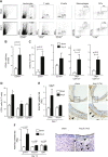
Leukocyte infiltration, chemokine receptors, and RANTES expression in pVAT during Ang II–dependent hypertension. Hypertension was induced by chronic 14 d Ang II infusion by osmotic minipump (490 ng/min/kg), and AT was obtained from periaortic fat pad (pVAT) and epididymal AT (visceral AT). A) Representative flow cytometric analysis of major leukocyte subpopulations in vascular stromal fraction isolated from periaortic AT of sham- and Ang II–infused mice. B) Effect of Ang II infusion on absolute numbers of CD45+ total leukocyte content in pVAT compartment expressed per mg of tissue (n = 14). C) Effect of Ang II infusion on content CD3+ T cells, CD19+ B cells, I-Ab+CD11b+ macrophages, and I-Ab+CD11c+ DCs in pVAT (n = 12–14 for each). D) Effect of Ang II–dependent hypertension on content of CCR1, CCR3, and CCR5+ T lymphocyte (CD3+) in isolated pVAT (n = 6). E) Effect of 7 d Ang II–induced hypertension on mRNA expression of RANTES in pVAT and visceral AT (n = 5), and immunostaining of aortas from sham-treated and Ang II–infused C57BL/6J mice using anti-RANTES antibody (representative of 5 experiments). F) Effect of 14 d Ang II–induced hypertension on mRNA expression of RANTES in pVAT, visceral AT, and BAT (n = 5) and immunostaining of aortas from sham-treated and Ang II–infused C57BL/6J mice using anti-RANTES antibody (representative of 5 experiments).
Figure 2.
Figure 2.
Role of RANTES in T lymphocyte migration in response to Ang II. Experiments were performed in modified Boyden chamber. A, B) Migration of T cells and their subsets and B cells (as reference) from sham- or Ang II–infused mice toward soluble RANTES (10 ng/ml, n = 9). C, D) Chemotaxis of T cells and CD4+ and CD8+ T cells toward conditioned medium from pVAT from sham- and Ang II–infused mice (n = 6 for each). E) Effects of anti-RANTES neutralizing antibody pretreatment on T-cell chemotaxis toward pVAT conditioned medium from sham- and Ang II–treated mice. Isotype antibody–pretreated medium was treated as 100%. Data are expressed as means ± sem.
Figure 3.
Figure 3.
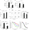
RANTES in Ang II–dependent hypertension and regulation of vascular dysfunction in animal model and in humans. A) Effect of Ang II–induced hypertension on endothelium-dependent vasodilatation to ACh in aortas of WT and RANTES−/− mice (n = 6 for each). B) Relaxations to sodium nitroprusside as measure of non-endothelium-dependent vasodilatation (n = 6 for each). Statistical analysis was performed by repeated measures ANOVA. C) Aortic superoxide levels measured by monitoring oxidation of dihydroethidium to 2-hydroxyethidium using HPLC in WT and RANTES−/− mice infused for 14 d with buffer (sham) or Ang II (n = 5 each group). D) Mean daily values of invasive telemetric measurements of systolic (top left), diastolic (bottom left ), and mean arterial (top right) blood pressure and heart rate (bottom right) at baseline and during Ang II infusion in WT and RANTES−/− mice (n = 6). E) Correlation between serum RANTES levels and FMD in high-cardiovascular-risk cohort of 129 subjects. F) Relationship between RANTES serum levels and non-endothelium-dependent nitroglycerin-mediated dilatation induced vasodilatation this cohort. G) Relationship between RANTES and vWF (as biochemical marker for endothelial dysfunction) levels in serum of high-cardiovascular-risk cohort. E–G) Statistics for these relationships presented as Spearman’s correlation tests.
Figure 4.
Figure 4.
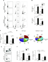
Role of RANTES in Ang II–dependent hypertension and T-cell perivascular infiltration. A) Examples of flow cytometric determination of effects of Ang II infusion on isolated pVAT (minus aorta) infiltration with total leukocytes (CD45+) and T cells (CD3+) in WT and RANTES−/− mice. B) Effect of Ang II–dependent hypertension on mean total leukocyte (CD45+ cells) and T-cell (CD3+) content in isolated pVAT in WT and RANTES−/− mice (n = 5 each). C) Effect of Ang II–dependent hypertension on mean macrophage infiltration in pVAT (n = 5 each). D) Differences in leukocyte subpopulation composition of pVAT upon Ang II infusion in WT and RANTES−/− mice showing notable reduction of T-cell content (n = 5 each). E) Gating strategy for detection of M2 (CD11cCD206+) and M1 type AT macrophages (CD11c+CD206) within F4/80+CD11b+ cells. F) Effect of Ang II–dependent hypertension on mean M1 (left) and M2 (right) macrophage infiltration in pVAT upon Ang II infusion in WT and RANTES−/− mice (n = 5 each).
Figure 5.
Figure 5.
T-cell subsets in isolated pVAT are regulated by RANTES in hypertension; links to vascular dysfunction. A, B) Flow cytometric analyses were used to determine number of CCR5+ T cells (A) and CCR6+ T cells (B) in pVAT of sham- and Ang II–infused mice (n = 5). C) Ang II–dependent changes in IL-17-producing CD4+ T cells in pVAT from WT and RANTES−/− mice (n = 5). D) Ang II–dependent changes in IFN-γ-producing CD8+ T cells in pVAT from WT and RANTES−/− mice (n = 5). E) Effect of Ang II on mRNA expression (real-time PCR) of IFN-γ in pVAT from WT and RANTES−/− mice (n = 5). F) Effects of IFN-γ (50 ng/ml) on endothelium-dependent and -independent relaxations in mouse aorta. Role of reactive oxygen species was examined using PEG-SOD (500 IU/ml) preincubation (n = 6; P, repeated measures ANOVA).
Figure 6.
Figure 6.
Double-negative CD3+CD4CD8 T cells are abundantly recruited to vasculature in Ang II–dependent hypertension and contribute to IFN-γ production. A) Effect of Ang II–dependent hypertension on mean CD3+CD4CD8 T-cell content in isolated pVAT in WT and RANTES−/− mice (n = 5 each; example of staining at left). B) Ang II–dependent changes in IFN-γ-producing CD3+CD4CD8 T cells in pVAT from WT and RANTES−/− mice (n = 5). Data are expressed as means ± sem.
Figure 7.
Figure 7.
Effects of met-RANTES on vascular function and perivascular T-cell infiltration in Ang II–dependent hypertension. A) Effect of Ang II–induced hypertension on endothelium-dependent vasodilatation to ACh in aortas of saline (saline) and met-RANTES-treated (50 mg/kg i.p.) mice (left; n = 5 for each). Relaxations to sodium nitroprusside as measure of non-endothelium-dependent vasodilatation (right; n = 5 for each). B) Aortic superoxide levels measured by monitoring oxidation of dihydroethidium to 2-hydroxyethidium using HPLC in control and met-RANTES-treated mice infused for 14 d with buffer (sham) or Ang II (n = 5). C) Effects of met-RANTES on mean T-cell (CD45+CD3+) infiltration in isolated pVAT (minus aorta) during Ang II–dependent hypertension (n = 5).

References

    1. Omar A., Chatterjee T. K., Tang Y., Hui D. Y., Weintraub N. L. (2014) Proinflammatory phenotype of perivascular adipocytes. Arterioscler. Thromb. Vasc. Biol. 34, 1631–1636 - PMC - PubMed
    1. Margaritis M., Antonopoulos A. S., Digby J., Lee R., Reilly S., Coutinho P., Shirodaria C., Sayeed R., Petrou M., De Silva R., Jalilzadeh S., Demosthenous M., Bakogiannis C., Tousoulis D., Stefanadis C., Choudhury R. P., Casadei B., Channon K. M., Antoniades C. (2013) Interactions between vascular wall and perivascular adipose tissue reveal novel roles for adiponectin in the regulation of endothelial nitric oxide synthase function in human vessels. Circulation 127, 2209–2221 - PubMed
    1. Gao Y. J. (2007) Dual modulation of vascular function by perivascular adipose tissue and its potential correlation with adiposity/lipoatrophy-related vascular dysfunction. Curr. Pharm. Des. 13, 2185–2192 - PubMed
    1. Wu H., Ghosh S., Perrard X. D., Feng L., Garcia G. E., Perrard J. L., Sweeney J. F., Peterson L. E., Chan L., Smith C. W., Ballantyne C. M. (2007) T-cell accumulation and regulated on activation, normal T cell expressed and secreted upregulation in adipose tissue in obesity. Circulation 115, 1029–1038 - PubMed
    1. Takemori K., Gao Y. J., Ding L., Lu C., Su L. Y., An W. S., Vinson C., Lee R. M. (2007) Elevated blood pressure in transgenic lipoatrophic mice and altered vascular function. Hypertension 49, 365–372 - PubMed

Publication types