Comparative Study
. 2017 Mar 1;27(3):1817-1830.
doi: 10.1093/cercor/bhw023.

Subtype-Specific Genes that Characterize Subpopulations of Callosal Projection Neurons in Mouse Identify Molecularly Homologous Populations in Macaque Cortex

Affiliations
Comparative Study

Subtype-Specific Genes that Characterize Subpopulations of Callosal Projection Neurons in Mouse Identify Molecularly Homologous Populations in Macaque Cortex

Ryann M Fame et al. Cereb Cortex. .

Abstract

Callosal projection neurons (CPN) interconnect the neocortical hemispheres via the corpus callosum and are implicated in associative integration of multimodal information. CPN have undergone differential evolutionary elaboration, leading to increased diversity of cortical neurons-and more extensive and varied connections in neocortical gray and white matter-in primates compared with rodents. In mouse, distinct sets of genes are enriched in discrete subpopulations of CPN, indicating the molecular diversity of rodent CPN. Elements of rodent CPN functional and organizational diversity might thus be present in the further elaborated primate cortex. We address the hypothesis that genes controlling mouse CPN subtype diversity might reflect molecular patterns shared among mammals that arose prior to the divergence of rodents and primates. We find that, while early expression of the examined CPN-enriched genes, and postmigratory expression of these CPN-enriched genes in deep layers are highly conserved (e.g., Ptn, Nnmt, Cited2, Dkk3), in contrast, the examined genes expressed by superficial layer CPN show more variable levels of conservation (e.g., EphA3, Chn2). These results suggest that there has been evolutionarily differential retraction and elaboration of superficial layer CPN subpopulations between mouse and macaque, with independent derivation of novel populations in primates. Together, these data inform future studies regarding CPN subpopulations that are unique to primates and rodents, and indicate putative evolutionary relationships.

Keywords: corpus callosum; development; evolution; primate; rodent.


Figures

Figure 1.
Figure 1.
Schematic representation and cytoarchitectural view of mouse and macaque somatosensory cortex. (A) Schematic representation of developing and adult mouse and macaque cortex in S1, drawn to a common internal scale. (B) DAPI nuclear stain at mid-corticogenesis (E14/ E94) and after neuronal migration (P3/ E108) in macaque to show cytoarchitectural layers in S1. Scale bars: B, 100 μm. E, embryonic day; P, postnatal day; VZ, ventricular zone; SVZ, subventricular zone; SP, subplate; ISVZ, inner subventricular zone; IFL, inner fiber layer; OSVZ, outer subventricular zone; OFL, outer fiber layer; CP, cortical plate; S1, primary somatosensory area; roman numerals indicate cortical layers. A, adapted from (Fame et al. 2011).
Figure 2.
Figure 2.
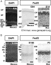
Fezf2 is detectable in macaque tissue using current in situ hybridization approach. (A) DAPI nuclear stain in E94 macaque for reference, followed by in situ hybridization for Fezf2 at E94 in macaque and E14 in mouse, showing early similarities in expression between mouse and macaque. (B) DAPI nuclear stain in E108 macaque for reference, followed by in situ hybridization for Fezf2 at E108 in macaque and P3 in mouse, confirming early similarities in expression between mouse and macaque, and validating the approach employed in these analyses. N = 3 individual mice, N = 2 technical replicates each. N = 1 macaque, N = 2 technical replicates. Scale bars: (A,B) 100 μm. E, embryonic day; P, postnatal day; VZ, ventricular zone; SVZ, subventricular zone; IZ, intermediate zone; CP, cortical plate; S1, primary somatosensory area; Roman numerals indicate cortical layers. E14 mouse in situ from GenePaint digital expression atlas www.genepaint.org.
Figure 3.
Figure 3.
At mid-corticogenesis (E14 and E94), early-expressed CPN genes are largely similarly expressed in mouse and macaque. (AH) in situ hybridization at E94 in macaque (A,A′,C,C′,E,E′,G,G′) and E14 in mouse (B,B′,D,D′,F,F′,H,H′) for early expressed CPN genes (Ptn, Lmo4, Inhba, Cited2) shows early similarities in expression between mouse and macaque. Low-level Inhba expression extends into progenitors in macaque, but not in mouse. N = 1 macaque, N = 3 technical replicates. Scale bar: 100 μm. E, embryonic day; P, postnatal day; VZ, ventricular zone; SVZ, subventricular zone; IZ, intermediate zone; CP, cortical plate; S1, primary somatosensory area; Roman numerals indicate cortical layers. E14 mouse in situ from GenePaint digital expression atlas www.genepaint.org.
Figure 4.
Figure 4.
At P3 in mouse, and at E108 and E113 in macaque, CPN genes reveal related populations in mouse and macaque superficial (Cited2, Chn2, Epha3, Limch1) and deep (Cited2) cortical layers. (AL) In situ hybridization at E108in macaque (A,D,G,J), at E113 in macaque (B,E,H,K), and at P3 in mouse (C,F,I,L), for Cited2 in layers II/III and V; and (AC), Chn2 (DF), Epha3 (GI), and Limch1 in superficial layers (JL) showing molecular similarities between cellular populations in mouse and macaque cortex. (A′–L′) detailed insets. N = 3 individual mice, N = 2 technical replicates each. N = 1 E108 macaque, N = 3 technical replicates. N= 1 E113 macaque, N = 2 technical replicates. Scale bars: (AL) 100 μm; (A′,B′,D′,E′,G′,G′,H′,J′,K′) 200 μm macaque; (C′,F′,I′,L′) 100 μm mouse. E, embryonic day; VZ, ventricular zone; SVZ, subventricular zone; IZ, intermediate zone; CP, cortical plate.
Figure 5.
Figure 5.
Some CPN genes have largely analogous developmental expression patterns in mouse and macaque, but with some divergence. (A) DAPI nuclear stain in E94 and E108 macaque for reference, horizontal bars indicate the extent of each named layer and are used to indicate dominant expression patterns in the following panels. (B) In situ hybridization at E94 in macaque and E14 in mouse for CPN genes with strong conservation of expression early in SVZ and CPN, as well as later at E108 and E113 (Dkk3, Nnmt) (C) in situ hybridization at E94 in macaque and E14 in mouse for Inhba shows early similarities in expression between mouse and macaque and more restricted expression later at E108 and E113. (D) In situ hybridization at E94 in macaque and E14 in mouse for Plxnd1 reveal divergence in early expression, but conservation later at E108 and E113 in macaque and P3 in mouse. (E) In situ hybridization for Gfra2 reveals that, while early expression is conserved, at later times gene expression in deep layers is conserved with additional expanded gene expression into layer II. N = 3 individual mice, N = 2 technical replicates each. N = 1 E108 macaque, N = 3 technical replicates. N= 1 E113 macaque, N = 2 technical replicates. Scale bars: 200 μm macaque; 100 μm mouse. E, embryonic day; P, postnatal day; VZ, ventricular zone; SVZ, subventricular zone; SP, subplate; ISVZ, inner subventricular zone; IFL, inner fiber layer; OSVZ, outer subventricular zone; OFL, outer fiber layer; CP, cortical plate; S1, primary somatosensory area; roman numerals indicate cortical layers. Horizontal gray bars indicate layers of dominant gene expression.
Figure 6.
Figure 6.
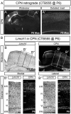
While superficial layer II/III does not contain the dominant proportion of CPN in mouse, superficial layer II/III CPN subpopulations do exist in P3 mouse cortex and express superficial layer II/III CPN genes, including Limch1. (A) Retrograde labeling in mouse using cholera toxin subunit B (CTB) reveals CPN cell bodies in cortical layer II/III at P8, including superficial portions of layer II/III. (B) In situ hybridization combined with retrograde labeling using CTB reveals CPN cell bodies in superficial portions of layer II/III whose localization overlaps with expression of the CPN gene Limch1 in mouse, suggesting that the layer II expression of Limch1 in Macaque (Fig. 4) might include CPN within that layer. (B′) Detailed medial inset from (B). (B″) Detailed lateral inset from (B). Scale bars: (B) 500 μm; (B′,B″) 100 μm. CPN, callosal projection neurons; CTB, B subunit of cholera toxin, P, postnatal day.
Figure 7.
Figure 7.
Functional areal and subcellular localization of CPN-expressed proteins suggests related functions in rodents and primates for conserved genes expressed by CPN populations. (A,A′,A″) Areal restriction of LMO4 is conserved between mouse and macaque. LMO4 expression in motor cortex extends throughout all neocortical layers, and more restricted expression limited to deep layers is observed in somatosensory cortex. (B,B′,C,C′,D,D′) Three proteins with distinct subcellular localization in mouse CPN were selected for study of subcellular localization in macaque: 1) Nectin-3, with axonal white matter localization by superficial layer CPN axons in the CC; 2) CAV1, with cell body and neurite localization by a subpopulation of deep layer CPN; and 3) LMO4, with nuclear localization in deep layer CPN of somatosensory cortex. (B,B′) At E94, Nectin-3 is localized specifically in white matter tracts in developing macaque cortex. There is low-level localization in the cell-dense cortical plate, higher level localization throughout the cell-sparse subplate, and very high-level fiber-localization in both the outer and inner fibrous layers. (C,C′) CAV1 is localized to neuronal cell membranes and neurites, as well as to developing blood vessels, as is seen in mouse ((Gaillard et al. 2001; Boulware et al. 2007; Molyneaux et al. 2009), and (D,D′) LMO4 shows nuclear localization in deep layer somatosensory neocortical neurons, as has been shown in mouse. Empty arrowheads indicate membranous localization, and filled arrows indicate nuclear localization. Scale bars: (A) 1 mm; and (A′,A″) 100 μm; (BD) 100 μm; (C′,D′) 50 μm. E, embryonic day; P, postnatal day; ICS, incipient central sulcus; PCG, postcentral gyrus; S1, primary somatosensory area; M1, primary motor area; VZ, ventricular zone; SVZ, subventricular zone; SP, subplate; ISVZ, inner subventricular zone; IFL, inner fiber layer; OSVZ, outer subventricular zone; OFL, outer fiber layer; CP, cortical plate; roman numerals indicate cortical layers.

Similar articles

Cited by

References

    1. Aboitiz F, Montiel J. 2003. One hundred million years of interhemispheric communication: the history of the corpus callosum. Braz J Med Biol Res. 36:409–420. - PubMed
    1. Aboitiz F, Morales D, Montiel J. 2003. The evolutionary origin of the mammalian isocortex: towards an integrated developmental and functional approach. Behav Brain Sci. 26:535–552; discussion 552–585. - PubMed
    1. Arcila ML, Betizeau M, Cambronne XA, Guzman E, Doerflinger N, Bouhallier F, Zhou H, Wu B, Rani N, Bassett DS et al. . 2014. Novel primate miRNAs coevolved with ancient target genes in germinal zone-specific expression patterns. Neuron. 81:1255–1262. - PMC - PubMed
    1. Arlotta P, Molyneaux BJ, Chen J, Inoue J, Kominami R, Macklis JD. 2005. Neuronal subtype-specific genes that control corticospinal motor neuron development in vivo. Neuron. 45:207–221. - PubMed
    1. Arnold SJ, Huang G-J, Cheung AFP, Era T, Nishikawa S-I, Bikoff EK, Molnár Z, Robertson EJ, Groszer M. 2008. The T-box transcription factor Eomes/Tbr2 regulates neurogenesis in the cortical subventricular zone. Genes Dev. 22:2479–2484. - PMC - PubMed

Publication types

MeSH terms