

Isolated omental metastasis of renal cell carcinoma after extraperitoneal open partial nephrectomy: A case report
- PMID: 26874583
- PMCID: PMC4802132
- DOI: 10.1016/j.ijscr.2016.02.008
Isolated omental metastasis of renal cell carcinoma after extraperitoneal open partial nephrectomy: A case report
Abstract
Introduction: Metachronous metastatic spread of clinically localized renal cell carcinoma (RCC) affects almost 1/3 of the patients. They occur most frequently in lung, liver, bone and brain. Isolated omental metastasis of RCC has not been reported so far.
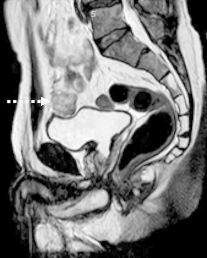
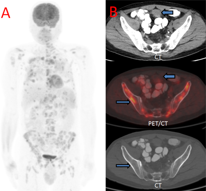
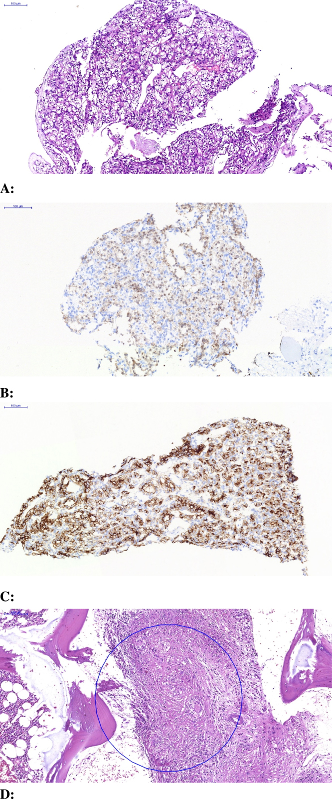
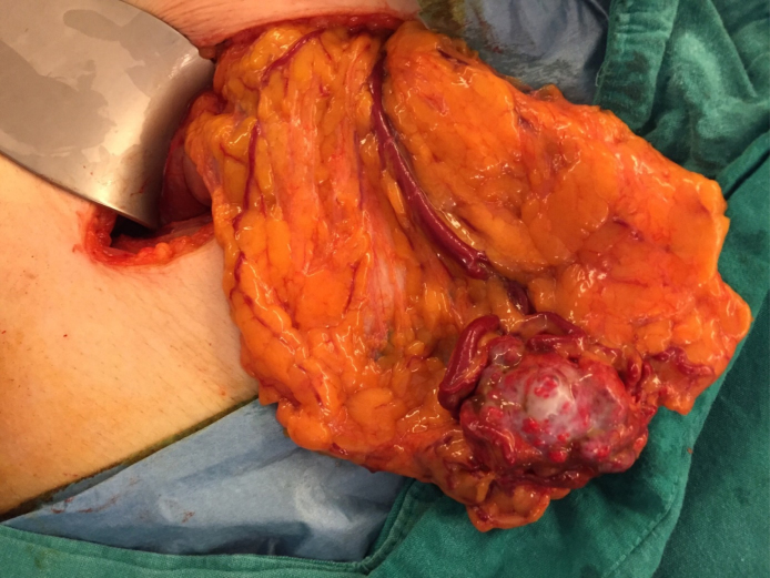
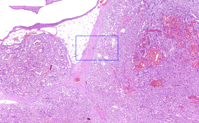
Case presentation: A 62-year-old patient previously diagnosed and treated due to pulmonary sarcoidosis has developed an omental metastatic lesion 13 years after having undergone open extraperitoneal partial nephrectomy for T1 clear-cell RCC. Constitutional symptoms and imaging findings that were attributed to the presence of a sarcomatoid paraneoplastic syndrome triggered by the development this metastatic focus complicated the diagnostic work-up. Biopsy of the [18F]-fluorodeoxyglucose (+) lesions confirmed the diagnosis of metastatic RCC and the patient was managed by the resection of the omental mass via near-total omentectomy followed by targeted therapy with a tyrosine kinase inhibitor.
Discussion: Late recurrence of RCC has been reported to occur in 10-20% of the patients within 20 years. Therefore lifelong follow up of RCC has been advocated by some authors. Diffuse peritoneal metastases have been reported in certain RCC subtypes with adverse histopathological features. However, isolated omental metastasis without any sign of peritoneal involvement is an extremely rare condition.
Conclusion: To our knowledge, this is the first reported case of metachronously developed, isolated omental metastasis of an initially T1 clear-cell RCC. Constitutional symptoms, despite a long interval since nephrectomy, should raise the possibility of a paraneoplastic syndrome being associated with metastatic RCC. Morphological and molecular imaging studies together with histopathological documentation will be diagnostic.
Keywords: Case report; Metastasis; Omentum; Renal cell carcinoma.
Copyright © 2016 The Authors. Published by Elsevier Ltd.. All rights reserved.
Figures
References
-
- Karumanchi S.A., Merchan J., Sukhatme V.P. Renal cancer: molecular mechanisms and newer therapeutic options. Curr. Opin. Nephrol. Hypertens. 2002;11(1):37–42. - PubMed
-
- Ljungberg B., Campbell S.C., Choi H.Y. The epidemiology of renal cellcarcinoma. Eur. Urol. 2011;60(4):615–621. - PubMed
-
- McNichols D.W., Segura J.W., DeWeerd J.H. Renal cell carcinoma: long-term survival and late recurrence. J. Urol. 1981;126:17–23. - PubMed
-
- Park Y.H., Baik K.D., Lee Y.J. Late recurrence of renal cell caricinoma >5 years after surgery: clinicopathological characteristics and prognosis. BJU Int. 2012 (Epub ahead of print) - PubMed
LinkOut - more resources
Full Text Sources
Other Literature Sources
