. 2016 Feb 29;49(4):550-7.
doi: 10.1016/j.jbiomech.2016.01.007. Epub 2016 Jan 14.

Human cartilage endplate permeability varies with degeneration and intervertebral disc site

Affiliations

Human cartilage endplate permeability varies with degeneration and intervertebral disc site

John F DeLucca et al. J Biomech. .

Abstract

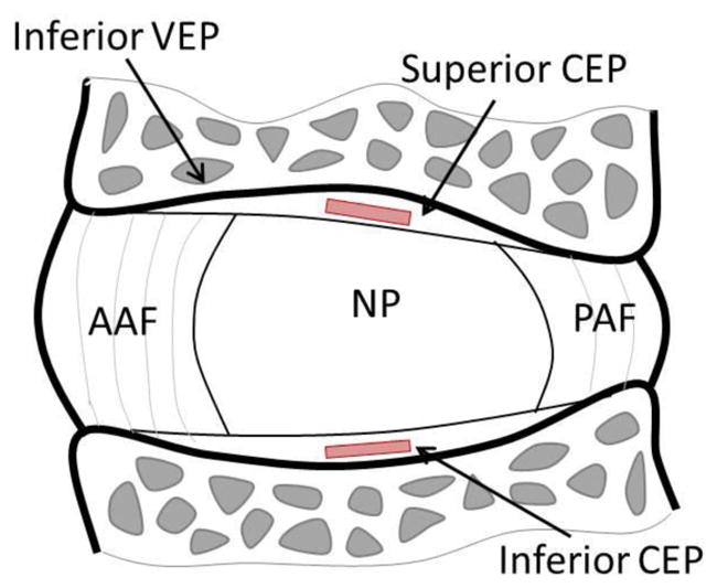
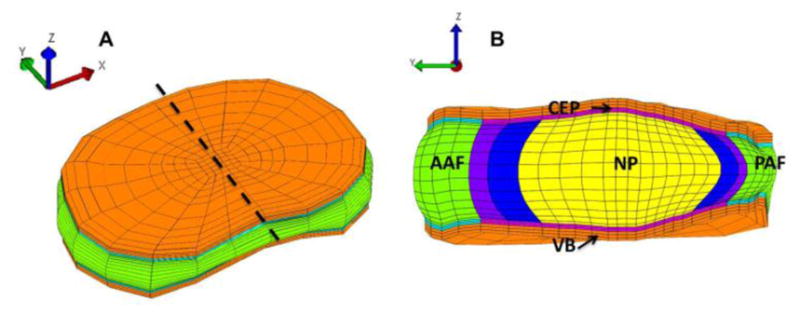
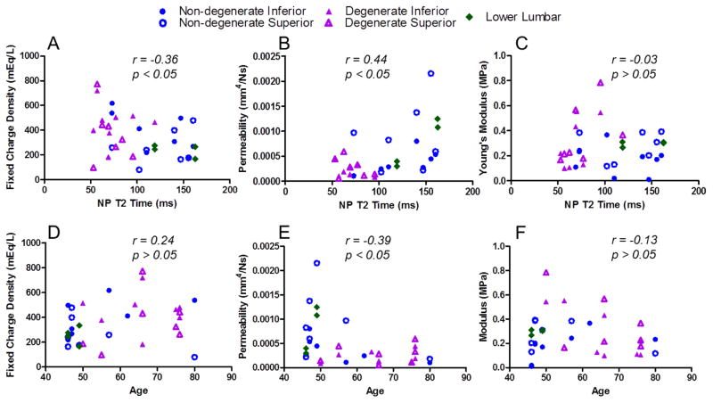
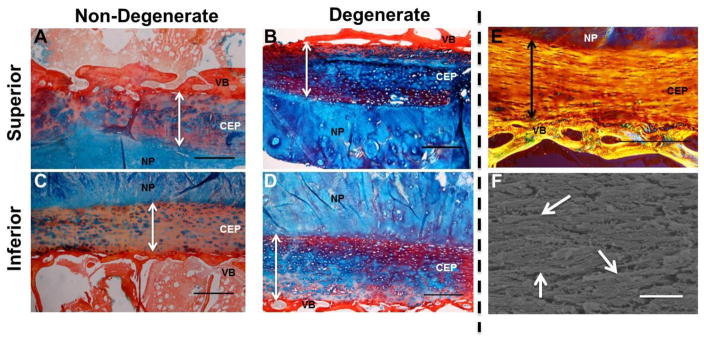
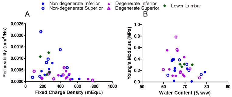
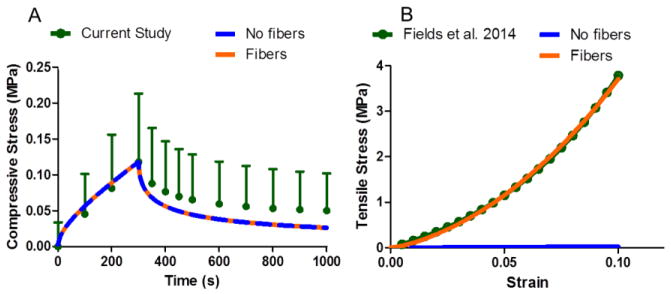
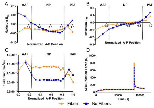
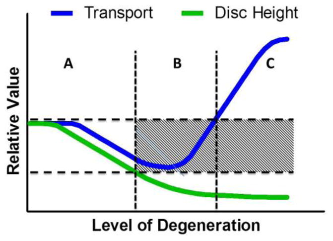
Despite the critical functions the human cartilage endplate (CEP) plays in the intervertebral disc, little is known about its structural and mechanical properties and their changes with degeneration. Quantifying these changes with degeneration is important for understanding how the CEP contributes to the function and pathology of the disc. Therefore the objectives of this study were to quantify the effect of disc degeneration on human CEP mechanical properties, determine the influence of superior and inferior disc site on mechanics and composition, and simulate the role of collagen fibers in CEP and disc mechanics using a validated finite element model. Confined compression data and biochemical composition data were used in a biphasic-swelling model to calculate compressive extrafibrillar elastic and permeability properties. Tensile properties were obtained by applying published tensile test data to an ellipsoidal fiber distribution. Results showed that with degeneration CEP permeability decreased 50-60% suggesting that transport is inhibited in the degenerate disc. CEP fibers are organized parallel to the vertebrae and nucleus pulposus and may contribute to large shear strains (0.1-0.2) and delamination failure of the CEP commonly seen in herniated disc tissue. Fiber-reinforcement also reduces CEP axial strains thereby enhancing fluid flux by a factor of 1.8. Collectively, these results suggest that the structure and mechanics of the CEP may play critical roles in the solute transport and disc mechanics.

Keywords: Biphasic; Cartilage endplate; Intervertebral disc; Permeability; Spine.


Conflict of interest statement

Conflicts of Interest

The authors have no conflicts of interest to report.

Figures

Fig. 1
Fig. 1
Sagittal section of the intervertebral disc showing location of CEP test samples taken adjacent to the nucleus pulposus (NP). CEP: cartilage endplate, AAF: anterior annulus fibrosus, PAF: posterior annulus fibrosus, NP: nucleus pulposus
Fig. 2
Fig. 2
Finite element model geometry and mesh shown for (A) the full disc, and (B) the mid-sagittal section, cut at the dashed line in (A). The full disc is oriented such that the anterior-posterior disc axis coincides with the y-axis. The CEP mechanics were calculated along the yz-plane. CEP: cartilage endplate, AAF: anterior annulus fibrosus, NP: nucleus pulposus, PAF: posterior annulus fibrosus, VB: vertebra
Fig. 3
Fig. 3
Mechanical parameters for all samples were evaluated for correlation with degeneration measured by nucleus pulposus (NP) T2 relaxation time and with specimen age. A lower T2 time represents a more degenerate disc. (A; D) Fixed charge density increases with degeneration (r = −0.36, p<0.05) and does not change with age (r = 0.24, p > 0.05) while (B; E) permeability decreases with degeneration (r = 0.44, p<0.05) and age (r = −0.39 , p < 0.05). (C; F) Modulus was not correlated with degeneration or age (p>0.05).
Fig. 4
Fig. 4
Histological assessment of non-degenerate and degenerate CEPs. Alcian blue and picrosirius red staining of superior (A,B) and inferior (C,D) endplates. Arrows denote CEP thickness. Non-degenerate CEP (A,C) appear structurally different than degenerate endplates (B, D). Non-degenerate CEP have GAG concentrated in the PCM (E) while degenerate CEP have prominent GAG staining throughout the tissue. Polarized light image viewed over 45° cross polarizers (E) and SEM image of CEP fibers (F). Fibers appear aligned in parallel to the vertebral bone and are not arranged like those found in articular cartilage. Arrows (F) denote example horizontally-oriented fibers. Scale bar (A–E) = 500 μm. Scale bar (F) = 1 μm. NP = Nucleus Pulposus, VB = Vertebra.
Fig. 5
Fig. 5
Permeability decreases with fixed charge density (r = −0.35, p<0.05; A) and modulus tended to decrease with water content (r = −0.31, p=0.08; B).
Fig. 6
Fig. 6
Fiber-reinforced model performance in compression and tension. Both models perform identically in confined compression (A) as fibers do not contribute in confined compression. However, fibers are essential to reproduce tensile data (B).
Fig. 7
Fig. 7
Compression stress-relaxation finite element results taken from the time step of maximum disc compression comparing the fiber-reinforced CEP model to the fiber-less model. AF and NP denote which disc sub-tissue is adjacent to the CEP with lines denoting tissue boundaries. Fibers significantly affect axial (A) and shear (B) deformations. Reduced CEP deformation allows for enhanced fluid flow (C). Fiber-reinforcement minimally affects global disc reaction force (D). CEP: cartilage endplate, AAF: anterior inner annulus fibrosus, PAF: posterior inner annulus fibrosus, NP: nucleus pulposus
Fig. 8
Fig. 8
Graph describing proposed changes in transport and disc height with degeneration. In non-degenerate discs, there is an unimpeded transport cycle (A). An optimal range of transport properties permits this balance. As degeneration proceeds, structural and biochemical changes to the CEP and disc reduce transport properties, inhibiting transport (B). In advanced degeneration, transport is enhanced but disc height substantially decreases (C).

References

    1. Antoniou J, Goudsouzian N, Heathfield T, Winterbottom N, Steffen T, Poole A, Aebi M, Alini M. The Human Lumbar Endplate. Spine (Phila Pa 1976) 1996;21:1153–1161. - PubMed
    1. Armstrong CG, V, Mow C. Variations in the intrinsic mechanical properties of human articular cartilage with age, degeneration, and water content. Journal of Bone and Joint Surgery. 1982;64:88–94. - PubMed
    1. Ateshian GA, Rajan V, Chahine NO, Canal CE, Hung CT. Modeling the matrix of articular cartilage using a continuous fiber angular distribution predicts many observed phenomena. Journal of biomechanical engineering. 2009;131:061003. - PMC - PubMed
    1. Ateshian GA, Soltz MA, Mauck RL, Basalo IM, Hung CT, Lai WM. The role of osmotic pressure and tension-compression nonlinearity in the frictional response of articular cartilage. Transport in Porous Media. 2003;50:5–33.
    1. Athanasiou KA, Rosenwasser MP, Buckwalter JA, Malinin TI, Mow VC. Interspecies comparisons of in situ intrinsic mechanical properties of distal femoral cartilage. Journal of Orthopedic Research. 1991;9:330–340. - PubMed

Publication types

LinkOut - more resources