

Prevalence of Subretinal Drusenoid Deposits in Older Persons with and without Age-Related Macular Degeneration, by Multimodal Imaging
- PMID: 26875000
- PMCID: PMC4842107
- DOI: 10.1016/j.ophtha.2015.12.034
Prevalence of Subretinal Drusenoid Deposits in Older Persons with and without Age-Related Macular Degeneration, by Multimodal Imaging
Abstract
Purpose: To assess the prevalence of subretinal drusenoid deposits (SDD) in older adults with healthy maculas and early and intermediate age-related macular degeneration (AMD) using multimodal imaging.
Design: Cross-sectional study.
Participants: A total of 651 subjects aged ≥60 years enrolled in the Alabama Study of Early Age-Related Macular Degeneration from primary care ophthalmology clinics.
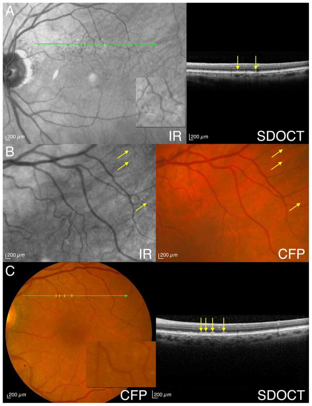
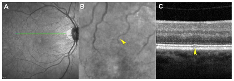
Methods: Subjects were imaged using spectral domain optical coherence tomography (SD OCT) of the macula and optic nerve head (ONH), infrared reflectance, fundus autofluorescence, and color fundus photographs (CFP). Eyes were assessed for AMD presence and severity using the Age-Related Eye Disease Study (AREDS) 9-step scale. Criteria for SDD presence were identification on ≥1 en face modality plus SD OCT or on ≥2 en face modalities if absent on SD OCT. Subretinal drusenoid deposits were considered present at the person level if present in 1 or both eyes.
Main outcome measures: Prevalence of SDD in participants with and without AMD.
Results: Overall prevalence of SDD was 32% (197/611), with 62% (122/197) affected in both eyes. Persons with SDD were older than those without SDD (70.6 vs. 68.7 years, P = 0.0002). Prevalence of SDD was 23% in subjects without AMD and 52% in subjects with AMD (P < 0.0001). Among those with early and intermediate AMD, SDD prevalence was 49% and 79%, respectively. After age adjustment, those with SDD were 3.4 times more likely to have AMD than those without SDD (95% confidence interval, 2.3-4.9). By using CFP only for SDD detection per the AREDS protocol, prevalence of SDD was 2% (12/610). Of persons with SDD detected by SD OCT and confirmed by at least 1 en face modality, 47% (89/190) were detected exclusively on the ONH SD OCT volume.
Conclusions: Subretinal drusenoid deposits are present in approximately one quarter of older adults with healthy maculae and in more than half of persons with early to intermediate AMD, even by stringent criteria. The prevalence of SDD is strongly associated with AMD presence and severity and increases with age, and its retinal topography including peripapillary involvement resembles that of rod photoreceptors. Consensus on SDD detection methods is recommended to advance our knowledge of this lesion and its clinical and biologic significance.
Copyright © 2016 American Academy of Ophthalmology. Published by Elsevier Inc. All rights reserved.
Conflict of interest statement
Conflict of Interest: no conflicting relationship exists for any author.
Figures

Comment in
-
Re: Zarubina et al.: Prevalence of subretinal drusenoid deposits in older persons with and without age-related macular degeneration, by multimodal imaging (Ophthalmology 2016;123:1090-1100).Ophthalmology. 2017 Feb;124(2):e19-e20. doi: 10.1016/j.ophtha.2016.05.014. Ophthalmology. 2017. PMID: 28126086 No abstract available.
-
Reply.Ophthalmology. 2017 Feb;124(2):e20-e21. doi: 10.1016/j.ophtha.2016.05.013. Ophthalmology. 2017. PMID: 28126088 Free PMC article. No abstract available.
References
-
- Schmitz-Valckenberg S, Alten F, Steinberg JS, et al. Reticular drusen associated with geographic atrophy in age-related macular degeneration. Invest Ophthalmol Vis Sci. 2011;52(9):5009–15. - PubMed
-
- Oak AS, Messinger JD, Curcio CA. Subretinal drusenoid deposits: further characterization by lipid histochemistry. Retina. 2014;34(4):825–6. - PubMed
Publication types
MeSH terms
Substances
Grants and funding
LinkOut - more resources
Full Text Sources
Other Literature Sources
Research Materials
Miscellaneous
