. 2016 Feb 15:6:21584.
doi: 10.1038/srep21584.

Constructing modular and universal single molecule tension sensor using protein G to study mechano-sensitive receptors

Affiliations

Constructing modular and universal single molecule tension sensor using protein G to study mechano-sensitive receptors

Xuefeng Wang et al. Sci Rep. .

Abstract

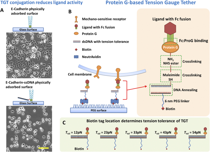
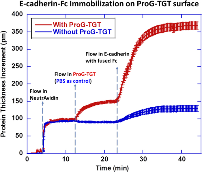
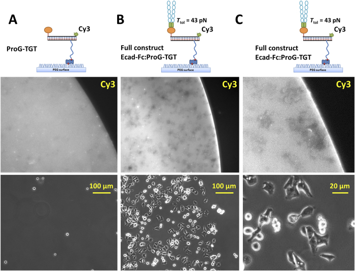
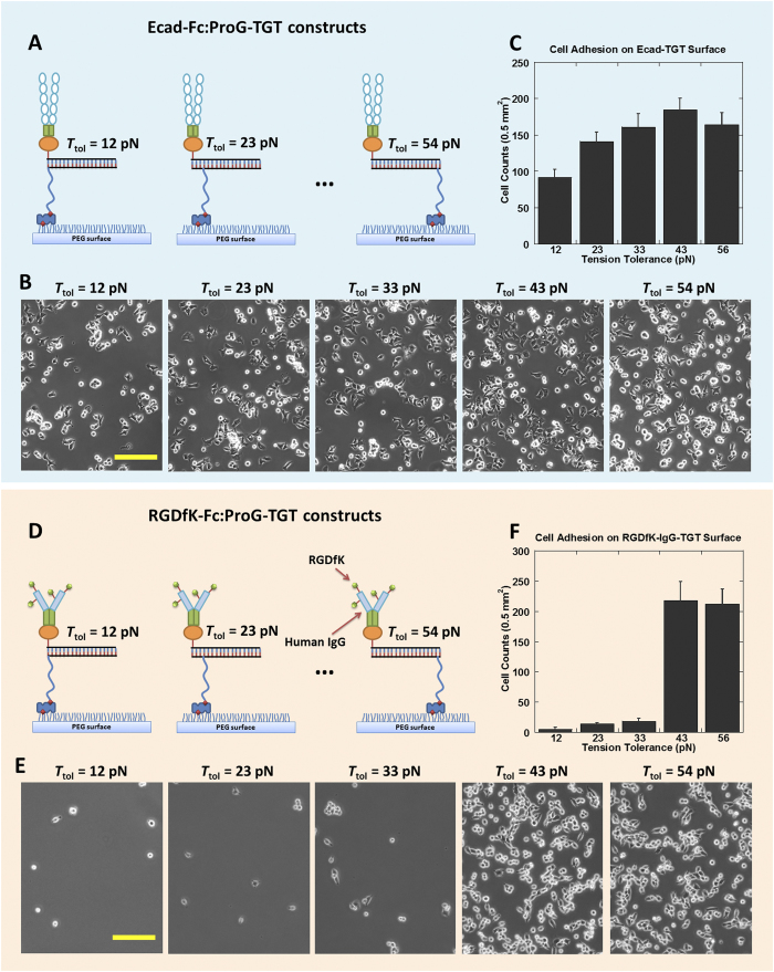
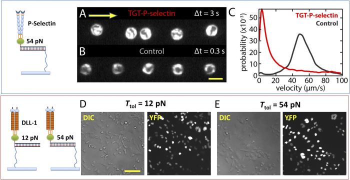
Recently a variety of molecular force sensors have been developed to study cellular forces acting through single mechano-sensitive receptors. A common strategy adopted is to attach ligand molecules on a surface through engineered molecular tethers which report cell-exerted tension on receptor-ligand bonds. This approach generally requires chemical conjugation of the ligand to the force reporting tether which can be time-consuming and labor-intensive. Moreover, ligand-tether conjugation can severely reduce the activity of protein ligands. To address this problem, we developed a Protein G (ProG)-based force sensor in which force-reporting tethers are conjugated to ProG instead of ligands. A recombinant ligand fused with IgG-Fc is conveniently assembled with the force sensor through ProG:Fc binding, therefore avoiding ligand conjugation and purification processes. Using this approach, we determined that molecular tension on E-cadherin is lower than dsDNA unzipping force (nominal value: 12 pN) during initial cadherin-mediated cell adhesion, followed by an escalation to forces higher than 43 pN (nominal value). This approach is highly modular and potentially universal as we demonstrate using two additional receptor-ligand interactions, P-selectin &PSGL-1 and Notch &DLL1.


Figures

Figure 1
Figure 1. ProG-based Tension Gauge Tether.
(A) Direct DNA conjugation on cadherin molecules reduced cadherin activity, leading to poorer DLD-1 cell adhesion and spreading. (B) The schematics of ligand immobilization through ProG-based TGT. Recombinant ligands with IgG-Fc fusion are assembled with ProG-TGT and immobilized on a glass surface passivated with polyethylene glycol (PEG). (C) Biotin tag was used to immobilize the ligand-TGT constructs. Biotin tag location on the dsDNA determines the tension tolerance Ttol of TGTs. Note that the Ttol values used in this article are nominal because the time scale of cellular force application is unknown. Scale bar: 100 μm.
Figure 2
Figure 2. Micro-ring resonator assay for immobilization of Fc-fused Ecad through ProG-TGT on polymer-passivated glass surface.
Concentration of reagents: 200 μg/mL neutravidin, 0.05 μM ProG-TGT and 50 μg/mL Ecad. Blue curve is the control run in which PBS buffer was flowed across the surface instead of ProG-TGT solution.
Figure 3
Figure 3. DLD-1 cells specifically adhere on Ecad-Fc:ProG-TGT coated surface.
(A) Cells did not adhere on ProG-TGT grafted surface (no Ecad-Fc). Middle panel shows a fluorescence image of Cy3 conjugated to ProG-TGT. The bright area marks the ProG-TGT coated region. Bottom panel shows a phase contrast image of the same area. (B) Cells specifically adhered on Ecad-Fc:ProG-TGT coated surface marked by strong fluorescence. Fluorescence loss (gray patches in TGT grafted area) underneath the cells suggests that cells caused TGT rupture. (C) Under higher magnification, it is evident that gray patches of fluorescence loss are co-localized with cells, confirming that TGTs were ruptured by DLD-1 cells.
Figure 4
Figure 4. Molecular tension measurement on cadherin and integrin.
(A) Schematics of Ecad immobilization through 12–54 pN ProG-based TGTs for cadherin tension measurement. (B) DLD-1 cells adhered on all 12~54 pN Ecad-Fc:ProG-TGT coated surfaces. (C) Cell adhesion density vs. Ttol. (D) Schematics of RGDfK-Fc immobilization through 12–54 pN ProG-based TGTs for integrin tension measurement. (E) Phase-contrast images of CHO-K1 cell adhesion on 12~54 pN RGDfK-Fc:ProG-TGT coated surfaces. (F) Cell adhesion density vs. Ttol. Scale bar: 100 μm.
Figure 5
Figure 5. ProG-TGT for P-selectin and Notch receptor.
(A) P-selectin-Fc:ProG-TGTs were assembled and immobilized on the inner surface of a flow chamber where leukocytes were flowed through. A single leukocyte location was captured by consecutive dark field imaging with 3 sec time interval. Yellow arrow indicates flow direction. (B) Single leukocyte location with 0.3 sec time interval on control surface without P-selectin coating. Scale bar: 20 μm. (C) Velocity distribution. HL-60 cells attached onto P-selectin-Fc:ProG-TGTs and show rolling behavior. This resulted in a cell moving rate significantly lower than the rate on control surface. (D) DLL1-Fc:ProG-TGTs were assembled and immobilized on glass surface where Notch activation was tested. Notch receptors were activated on both Ttol = 12 pN surface and Ttol = 54 pN surface after incubation for two days. Notch activation was reported by H2B-YFP expression in CHO-K1 cell nucleus. Scale bar: 50 μm.

References

    1. Giancotti F. G. & Ruoslahti E. Transduction - Integrin signaling. Science 285, 1028–1032 (1999). - PubMed
    1. Takeichi M. Cadherin Cell-Adhesion Receptors as a Morphogenetic Regulator. Science 251, 1451–1455 (1991). - PubMed
    1. Moore K. L. et al. P-Selectin Glycoprotein Ligand-1 Mediates Rolling of Human Neutrophils on P-Selectin. J Cell Biol 128, 661–671 (1995). - PMC - PubMed
    1. Artavanis-Tsakonas S., Rand M. D. & Lake R. J. Notch signaling: Cell fate control and signal integration in development. Science 284, 770–776 (1999). - PubMed
    1. Salaita K. et al. Restriction of Receptor Movement Alters Cellular Response: Physical Force Sensing by EphA2. Science 327, 1380–1385 (2010). - PMC - PubMed

Publication types

LinkOut - more resources