

doi: 10.1016/j.tim.2016.01.010.
Epub 2016 Feb 12.
A Deadly Path: Bacterial Spread During Bubonic Plague
Affiliations
- PMID: 26875618
- PMCID: PMC4808365
- DOI: 10.1016/j.tim.2016.01.010
Item in Clipboard
A Deadly Path: Bacterial Spread During Bubonic Plague
Trends Microbiol.
2016 Apr.
Abstract
Yersinia pestis causes bubonic plague, a fulminant disease where host immune responses are abrogated. Recently developed in vivo models of plague have resulted in new ideas regarding bacterial spread in the body. Deciphering bacterial spread is key to understanding Y. pestis and the immune responses it encounters during infection.
Keywords: Yersinia pestis; bubonic plague; dissemination.
Copyright © 2016 Elsevier Ltd. All rights reserved.
Figures
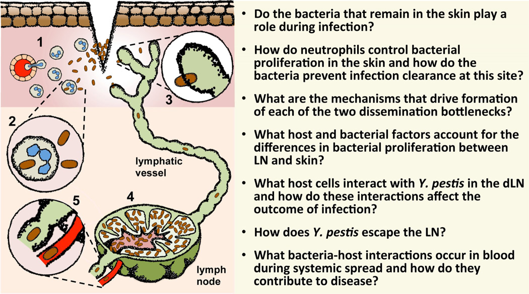
(1) Inoculation of Y. pestis (brown rods) into the dermal layer of the skin triggers the recruitment of neutrophils (light green cells with blue nuclei). The dermis (pink) appears bellow the epidermis (brown cuboidal cells). (2) In the dermis, Y. pestis interacts mostly with neutrophils. These cells control Y. pestis proliferation in the dermis but are unable to eliminate infection. (3) Dissemination competent bacteria escape from the skin immediately after inoculation via lymphatic vessels (green) to reach the draining lymph node. (4) Y. pestis colonizes the lymph node at very high rates. (5) The bacteria escape the lymph node compartment into systemic circulation through efferent lymphatic vessels or via blood vessels (red). The questions at the right of the figure summarize areas of the field that need to be explored to obtain a more detailed picture of how Y. pestis disseminates in the body.
Similar articles
-
Transcriptomic and innate immune responses to Yersinia pestis in the lymph node during bubonic plague.Infect Immun. 2010 Dec;78(12):5086-98. doi: 10.1128/IAI.00256-10. Epub 2010 Sep 27. Infect Immun. 2010. PMID: 20876291 Free PMC article.
-
Yersinia pestis YopJ suppresses tumor necrosis factor alpha induction and contributes to apoptosis of immune cells in the lymph node but is not required for virulence in a rat model of bubonic plague.Infect Immun. 2006 Sep;74(9):5126-31. doi: 10.1128/IAI.00219-06. Infect Immun. 2006. PMID: 16926404 Free PMC article.
-
Yersinia pestis subverts the dermal neutrophil response in a mouse model of bubonic plague.mBio. 2013 Aug 27;4(5):e00170-13. doi: 10.1128/mBio.00170-13. mBio. 2013. PMID: 23982068 Free PMC article.
-
Yersinia pestis and pneumonic plague: Insight into how a lethal pathogen interfaces with innate immune populations in the lung to cause severe disease.Cell Immunol. 2024 Sep-Oct;403-404:104856. doi: 10.1016/j.cellimm.2024.104856. Epub 2024 Jul 10. Cell Immunol. 2024. PMID: 39002222 Review.
-
Yersinia pestis and the plague.Am J Clin Pathol. 2003 Jun;119 Suppl:S78-85. doi: 10.1309/DQM9-3R8Q-NQWB-FYU8. Am J Clin Pathol. 2003. PMID: 12951845 Review.
Cited by
-
Proteogenomic discovery of sORF-encoded peptides associated with bacterial virulence in Yersinia pestis.Commun Biol. 2021 Nov 2;4(1):1248. doi: 10.1038/s42003-021-02759-x. Commun Biol. 2021. PMID: 34728737 Free PMC article.
-
Autophagy and Intracellular Membrane Trafficking Subversion by Pathogenic Yersinia Species.Biomolecules. 2020 Dec 4;10(12):1637. doi: 10.3390/biom10121637. Biomolecules. 2020. PMID: 33291818 Free PMC article. Review.
-
Dangerous Pathogens as a Potential Problem for Public Health.Medicina (Kaunas). 2020 Nov 6;56(11):591. doi: 10.3390/medicina56110591. Medicina (Kaunas). 2020. PMID: 33172013 Free PMC article. Review.
-
Bacterial Lymphatic Metastasis in Infection and Immunity.Cells. 2021 Dec 23;11(1):33. doi: 10.3390/cells11010033. Cells. 2021. PMID: 35011595 Free PMC article. Review.
-
PsaF Is a Membrane-Localized pH Sensor That Regulates psaA Expression in Yersinia pestis.J Bacteriol. 2021 Jul 22;203(16):e0016521. doi: 10.1128/JB.00165-21. Epub 2021 Jul 22. J Bacteriol. 2021. PMID: 34060904 Free PMC article.
References
MeSH terms
Grants and funding
LinkOut - more resources
Full Text Sources
Other Literature Sources
Medical
