. 2016 Feb 25;164(5):950-61.
doi: 10.1016/j.cell.2016.01.039. Epub 2016 Feb 11.

Structure and Engineering of Francisella novicida Cas9

Affiliations

Structure and Engineering of Francisella novicida Cas9

Hisato Hirano et al. Cell. .

Abstract

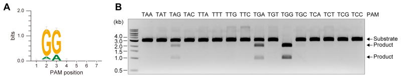
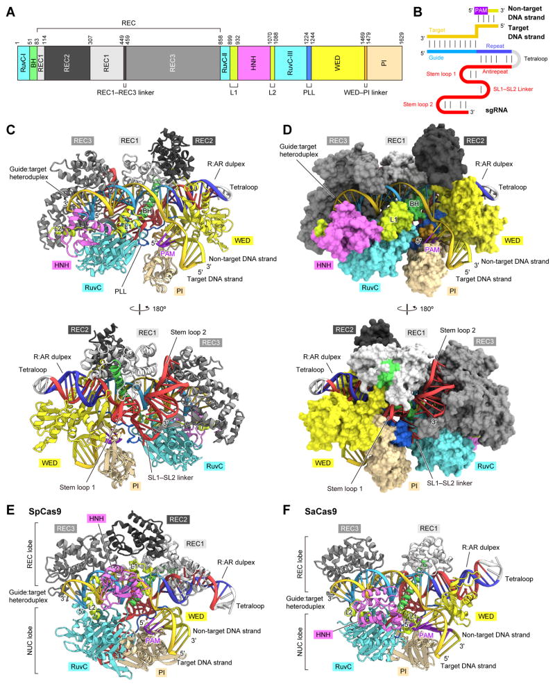
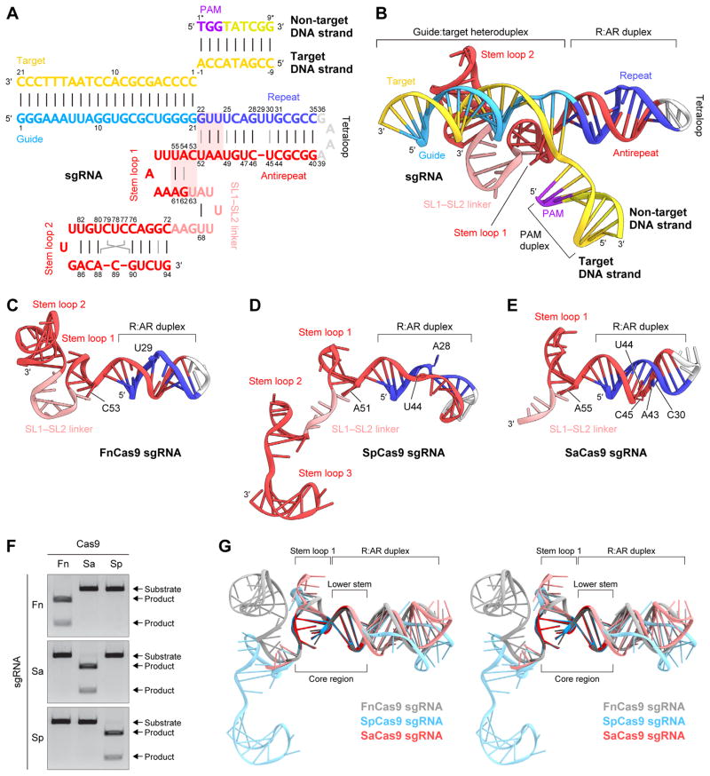
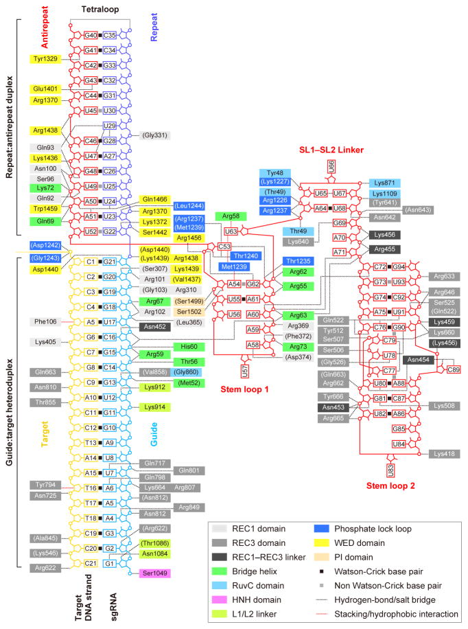
The RNA-guided endonuclease Cas9 cleaves double-stranded DNA targets complementary to the guide RNA and has been applied to programmable genome editing. Cas9-mediated cleavage requires a protospacer adjacent motif (PAM) juxtaposed with the DNA target sequence, thus constricting the range of targetable sites. Here, we report the 1.7 Å resolution crystal structures of Cas9 from Francisella novicida (FnCas9), one of the largest Cas9 orthologs, in complex with a guide RNA and its PAM-containing DNA targets. A structural comparison of FnCas9 with other Cas9 orthologs revealed striking conserved and divergent features among distantly related CRISPR-Cas9 systems. We found that FnCas9 recognizes the 5'-NGG-3' PAM, and used the structural information to create a variant that can recognize the more relaxed 5'-YG-3' PAM. Furthermore, we demonstrated that the FnCas9-ribonucleoprotein complex can be microinjected into mouse zygotes to edit endogenous sites with the 5'-YG-3' PAM, thus expanding the target space of the CRISPR-Cas9 toolbox.


Figures

Figure 1
Figure 1. PAM specificity of FnCas9
(A) PAM discovery assay for FnCas9. (B) In vitro DNA cleavage by FnCas9. The linearized plasmid targets with the 5′-TNN-3′ PAM were incubated with the purified FnCas9–sgRNA complex.
Figure 2
Figure 2. Overall structure of the FnCas9–sgRNA–DNA complex
(A) Domain organization of FnCas9. BH, bridge helix; PLL, phosphate lock loop. (B) Schematic representation of the sgRNA–DNA. (C and D) Ribbon (C) and surface (D) representations of the FnCas9–sgRNA–DNA complex. (E and F) Crystal structures of SpCas9 (PDB: 4UN3) (E) and SaCas9 (PDB: 5CZZ) (F).
Figure 3
Figure 3. Structure of sgRNA–DNA
(A) Schematic representation of the FnCas9 sgRNA scaffold. The sgRNA core fold is highlighted in pink. (B) Structure of the FnCas9 sgRNA–DNA. (C–E) sgRNA scaffolds for FnCas9 (C), SpCas9 (D) and SaCas9 (E). The guide regions are omitted for clarity. (F) Comparison of the sgRNA scaffolds of FnCas9 (red), SpCas9 (blue) and SaCas9 (gray).
Figure 4
Figure 4. Schematic of the nucleic acid recognition by FnCas9
Residues that interact with nucleic acids via their main chain are shown in parentheses. Water-mediated hydrogen bonds are not shown for clarity.
Figure 5
Figure 5. Recognition of the sgRNA and the target DNA by the Cas9 orthologs
(A–C) Recognition of the nucleic acids by the REC/WED domains of SpCas9 (A), SaCas9 (B) and FnCas9 (C). (D) Recognition of the RNA–DNA heteroduplex by FnCas9. Hydrogen-bonding and electrostatic interactions are indicated by gray dashed lines. (E) Recognition of the sgRNA core fold by FnCas9.
Figure 6
Figure 6. PAM recognition
(A) Binding of the PAM duplex to the groove between the WED and PI domains. (B) Schematics of the PAM duplex recognition. Water-mediated hydrogen bonds between the protein and the sugar-phosphate backbone are omitted for clarity. (C and D) Recognition of the 5′-TGG-3′ (C) and 5′-TGA-3′ (D) PAMs. Water molecules are shown as red spheres. (E) In vitro mutational analysis of the PAM-interacting residues. The linearized plasmid targets with the 5′-TGN-3′ PAMs were incubated with the wild type and mutants of FnCas9. (F) Comparison of the PI domains of SpCas9 (F), SaCas9 (G) and FnCas9 (H)
Figure 7
Figure 7. Structure-guided engineering and genome editing in mouse zygotes
(A) In vitro cleavage activity of wild-type and RHA FnCas9. The linearized plasmid targets with the 5′-TGN-3′ PAMs were incubated with the purified FnCas9–sgRNA complex. (B) PAM discovery assay for RHA FnCas9. (C) Preference of wild-type and RHA FnCas9 for the 1st PAM nucleotides. The linearized plasmid targets with the 5′-NGG-3′ PAMs were incubated with the FnCas9–sgRNA complex. (D) PAM recognition mechanism of wild-type (left) and RHA (right) FnCas9. (E) FnCas9-mediated genome editing in mouse zygotes. The pre-assembled wild-type and RHA FnCas9 RNP complexes were microinjected into mouse zygotes. The ratios between the numbers of embryos with FnCas9-mediated indels and the total numbers of injected embryos are shown above the bars. The numbers of embryos with mutations in both alleles (left) and a single allele (right) are shown in parentheses.

References

    1. Adams PD, Afonine PV, Bunkoczi G, Chen VB, Davis IW, Echols N, Headd JJ, Hung LW, Kapral GJ, Grosse-Kunstleve RW, et al. PHENIX: a comprehensive Python-based system for macromolecular structure solution. Acta Crystallogr D Biol Crystallogr. 2010;66:213–221. - PMC - PubMed
    1. Anders C, Niewoehner O, Duerst A, Jinek M. Structural basis of PAM-dependent target DNA recognition by the Cas9 endonuclease. Nature. 2014;513:569–573. - PMC - PubMed
    1. Briner AE, Donohoue PD, Gomaa AA, Selle K, Slorach EM, Nye CH, Haurwitz RE, Beisel CL, May AP, Barrangou R. Guide RNA functional modules direct Cas9 activity and orthogonality. Mol Cell. 2014;56:333–339. - PubMed
    1. Chylinski K, Le Rhun A, Charpentier E. The tracrRNA and Cas9 families of type II CRISPR-Cas immunity systems. RNA Biol. 2013;10:726–737. - PMC - PubMed
    1. Cong L, Ran FA, Cox D, Lin S, Barretto R, Habib N, Hsu PD, Wu X, Jiang W, Marraffini LA, et al. Multiplex genome engineering using CRISPR/Cas systems. Science. 2013;339:819–823. - PMC - PubMed

Publication types

Substances