Review
. 2016 Jun 8;116(11):6503-15.
doi: 10.1021/acs.chemrev.5b00590. Epub 2016 Feb 15.

Protein Allostery and Conformational Dynamics

Affiliations
Review

Protein Allostery and Conformational Dynamics

Jingjing Guo et al. Chem Rev. .

Abstract

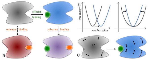
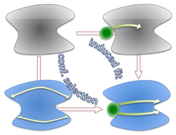
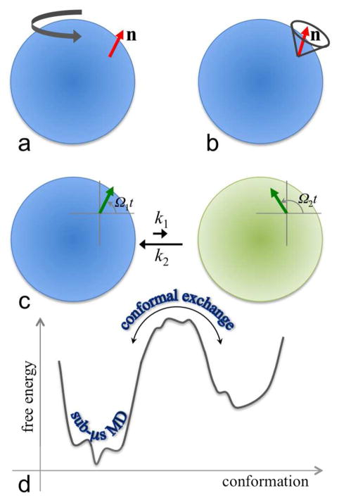
The functions of many proteins are regulated through allostery, whereby effector binding at a distal site changes the functional activity (e.g., substrate binding affinity or catalytic efficiency) at the active site. Most allosteric studies have focused on thermodynamic properties, in particular, substrate binding affinity. Changes in substrate binding affinity by allosteric effectors have generally been thought to be mediated by conformational transitions of the proteins or, alternatively, by changes in the broadness of the free energy basin of the protein conformational state without shifting the basin minimum position. When effector binding changes the free energy landscape of a protein in conformational space, the change affects not only thermodynamic properties but also dynamic properties, including the amplitudes of motions on different time scales and rates of conformational transitions. Here we assess the roles of conformational dynamics in allosteric regulation. Two cases are highlighted where NMR spectroscopy and molecular dynamics simulation have been used as complementary approaches to identify residues possibly involved in allosteric communication. Perspectives on contentious issues, for example, the relationship between picosecond-nanosecond local and microsecond-millisecond conformational exchange dynamics, are presented.


Figures

Figure 1
Figure 1
Conformational and dynamic effects of allosteric binding. (a) Binding of an effector at the allosteric site primes the binding of the substrate at the active site, and consequently the thermodynamic or kinetic properties of the latter binding are different from those in the absence of the effector. (b) Allosteric binding may result in a change in (Left) the conformational state, as signified by the movement of the corresponding free energy basin from one region to another region in conformational space, or (Right) the broadness of the free energy basin. (c) Conformation dynamics may be changed as well, e.g., from uncorrelated, fast (e.g., sub-nanosecond) motions in the apo form to correlated, slow (e.g., > microsecond) motions in the effector-bound form. Internal motions are represented by double-headed arrows.
Figure 2
Figure 2
Transition pathways connecting the end states of allosteric binding and communication pathways from the allosteric site to the active site. The allosteric transition illustrated here is a conformational change, with “apo” and “bound” conformations represented by gray and blue shading, respectively. Transition pathways, indicated by brown arrows, are mainly concerned with kinetic intermediates, with induced fit passing through the intermediate in which the effector is loosely bound with the protein in the apo conformation, whereas conformational selection passing through the intermediate in which the apo protein adopts the bound conformation. Communication pathways, indicated by green arrows, are concerned with intermediate residues through which the allosteric site is coupled with the active site. The two types of pathways thus differ in their emphases, but are not orthogonal to each other. In the induced-fit transition pathway, the conformational change initiated by loose effector binding propagates to the active site while the effector consolidates its binding. In the conformational-selection pathway, the stabilization of the bound conformation starts at the allosteric site and propagates to the active site.
Figure 3
Figure 3
Different types of conformational dynamics. (a) Overall rotational diffusion. The unit vector n is assumed to be rigidly attached to the protein represented by a sphere with blue shading. (b) Local orientational dynamics of n, illustrated here as diffusion in a cone, in the body-fixed frame. (c) Conformational exchange between a major state (shaded blue) and a minor state (shaded green). The magnetization (green arrow) precesses at different frequencies in the two states, resulting in dephasing. (d) Conformational sampling on different timescales. Sub-microsecond molecular dynamics simulations sample local fluctuations within a single conformational state (or substate therein). Transitions between conformational states require non-local correlated motions and cross high free energy barriers, typically occurring on the microsecond-millisecond timescale.
Figure 4
Figure 4
A protein represented as a weighted graph. (a) Shortest path between two nodes 1 and 7, composed of three connected edges shown in blue. Each edge (indicated by a line between two nodes) is assigned a path length (e.g., eq 16 or 17). The path lengths of the three blue edges are shown. Summing over the individual path lengths, the total length of the shortest path is 3.0. (b) The nodes are partitioned into two communities, one with green nodes inside a lime oval and one with red nodes inside a pink oval. An edge between nodes 5 and 6 links the two communities; these nodes are known as critical nodes.
Figure 5
Figure 5
Structure and allosteric communication of Pin1. (a) Structure of Pin1 with FFpSPR bound at the WW site. The catalytic site of the PPIase domain is lined by the three loops labeled as catalytic loop, β5-α4, and β6-β7. (b) Two clusters of paths connecting the WW domain to the catalytic-site loops. The cluster shown as light blue arrows preexists in apo Pin1, but the paths in the second cluster, shown as pink arrows, are broken in the apo form and are completed only in the FFpSPR-bound form. (c, d) Community analysis results for apo and FFpSPR-bound Pin1. The communities are shown in different colors as cartoon structures (Left) or as ovals (Middle). Intercommunity connections are shown as lines, with width proportional to the cumulative betweenness of inter-community edges (Middle). Shown in (Right) is a dynamic model for allostery. A spring depicts a representative internal coordinate from each of communities 1, 2, and 3 that is modeled as undergoing diffusive motion in a harmonic potential. The internal coordinates are weakly coupled in apo Pin1 and become strongly coupled in the FFpSPR-bound form.
Figure 6
Figure 6
Structure of IGPS. SideL and sideR are in light orange and magenta, respectively, for of HisF, and in gray and green, respectively for HisH. HisF sideL consists of residues 101-220, and HisH sideR consists of β1-β4 strands, α1, α2, α2′, and α4 helices, and Ω-loop. The bound PRFAR in HisF is shown in cyan spheres, and the catalytic triad and PGVG motif are labeled, as are some secondary structure elements (α1-α3 and loop1 in HisF and α1, α2, α2′, and Ω-loop in HisH).

References

    1. Monod J, Jacob F. General Conclusions: Telenomic Mechanisms in Cellular Metabolism, Growth, and Differentiation. Cold Spring Harbor Symp Quant Biol. 1961;26:389–401. - PubMed
    1. Monod J, Wyman J, Changeux JP. On the Nature of Allosteric Transitions: A Plausible Model. J Mol Biol. 1965;12:88–118. - PubMed
    1. Wyman J, Allen DW. The Problem of the Heme Interactions in Hemoglobin and the Basis of the Bohr Effect. J Polym Sci. 1951;7:499–518.
    1. Cooper A, Dryden DT. Allostery without Conformational Change. A Plausible Model. Eur Biophys J. 1984;11:103–109. - PubMed
    1. Nussinov R, Tsai CJ. Allostery without a Conformational Change? Revisiting the Paradigm. Curr Opin Struct Biol. 2015;30:17–24. - PubMed

Publication types

MeSH terms

Substances

LinkOut - more resources