. 2016 Mar 1;24(3):412-22.
doi: 10.1016/j.str.2015.12.017. Epub 2016 Feb 11.

Selective Binding of AIRAPL Tandem UIMs to Lys48-Linked Tri-Ubiquitin Chains

Affiliations

Selective Binding of AIRAPL Tandem UIMs to Lys48-Linked Tri-Ubiquitin Chains

Simin Rahighi et al. Structure. .

Abstract

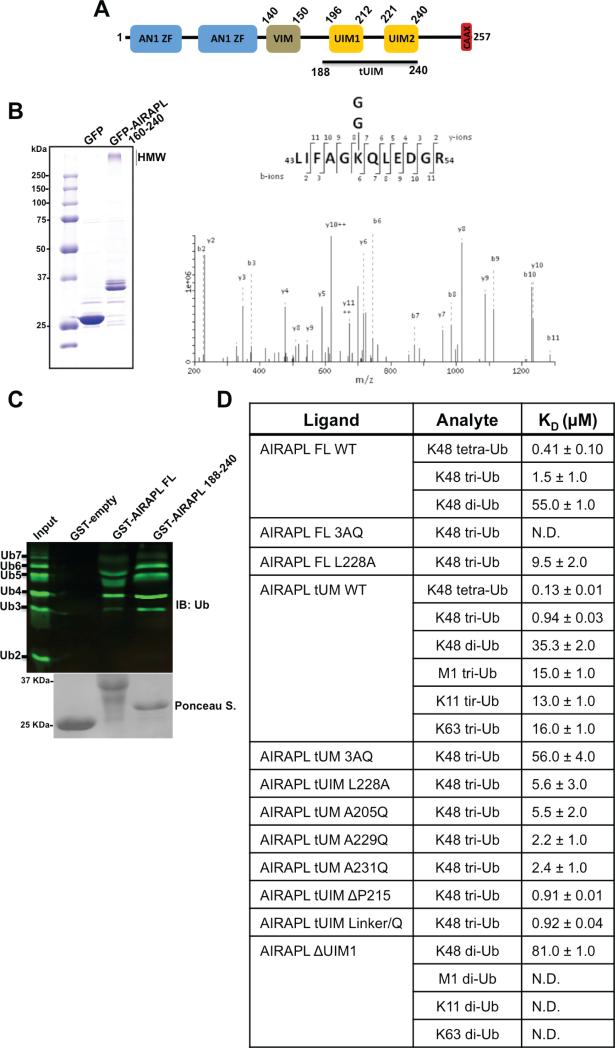
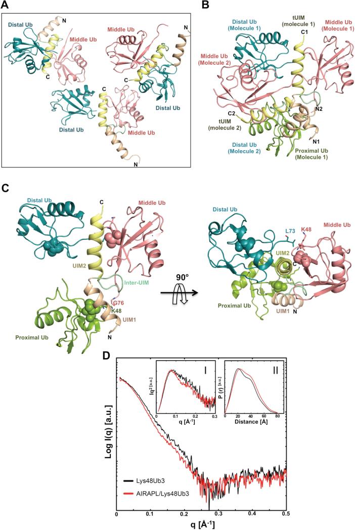
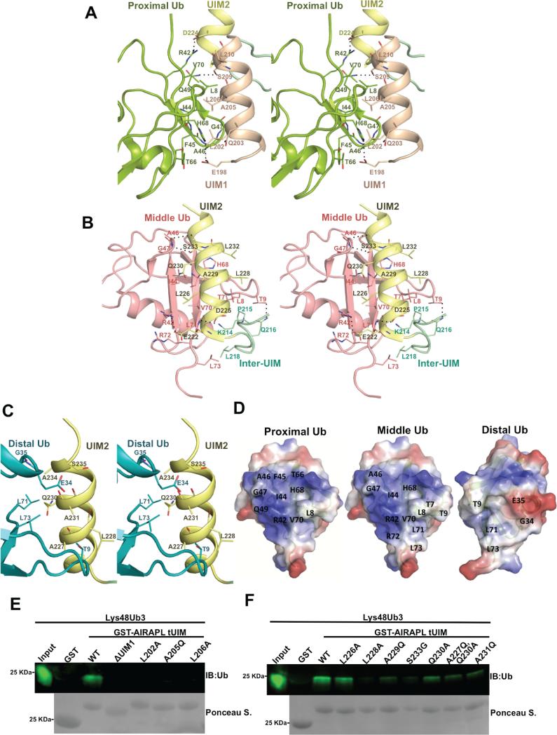
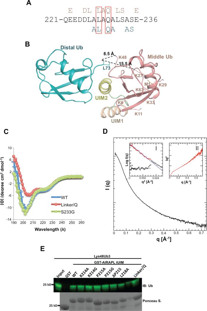
Lys48-linked ubiquitin chains act as the main targeting signals for protein degradation by the proteasome. Here we report selective binding of AIRAPL, a protein that associates with the proteasome upon exposure to arsenite, to Lys48-linked tri-ubiquitin chains. AIRAPL comprises two ubiquitin-interacting motifs in tandem (tUIMs) that are linked through a flexible inter-UIM region. In the complex crystal structure UIM1 binds the proximal ubiquitin, whereas UIM2 (the double-sided UIM) binds non-symmetrically to the middle and distal ubiquitin moieties on either side of the helix. Specificity of AIRAPL for Lys48-linked ubiquitin chains is determined by UIM2, and the flexible inter-UIM linker increases avidity by placing the two UIMs in an orientation that facilitates binding of the third ubiquitin to UIM1. Unlike middle and proximal ubiquitins, distal ubiquitin binds UIM2 through a novel surface, which leaves the Ile44 hydrophobic patch accessible for binding to the proteasomal ubiquitin receptors.


Figures

Figure 1
Figure 1. AIRAPL is an ubiquitin-binding protein and is selective for Lys48-linked ubiquitin chains
(A) Schematic domain organization of mouse AIRAPL protein. AN1 ZF, AN1-type zinc finger; VIM, valosin-containing protein (VCP)-interacting motif; UIM, ubiquitin-interacting motif; CAAX box, a sequence of cysaliphatic-aliphatic-undefined amino acids. (B) Left: high molecular weight (HMW) polyubiquitin chains co-purified with GFP tagged-AIRAPL (residues 160-240) were subjected to liquid chromatography-tandem mass spectrometry (LCMS/MS). GFP alone was used as a negative control for binding to ubiquitin. Right: the MS/MS spectrum of the Lys48-linkage GG-containing peptide 43-54 derived from Ub with a precursor ion mass of [M+2H] 2+ 731.0 Da was obtained from cells expressing Flag-AIRAPL. The b/y fragment ions are indicated. (C) Binding of GST-tagged AIRAPL FL and tUIM to Lys48-linked (2-7)-ubiquitin chains was analyzed by immunoblotting using anti-ubiquitin antibody. Loading of GST-tagged proteins was determined by Ponceau S staining. (See also Fig. S1) (D) Binding affinity (KD or equilibrium constants) of AIRAPL FL, tUIM and ΔUIM1, WT (wild type) or mutants, for various ubiquitin chain types and lengths measured by surface plasmon resonance (SPR). For each measurement, His-tagged AIRAPL was immobilized on NiHC1000m chip surface and ubiquitin chains were loaded over the chip. Each measurement was done at least two times and data was analyzed using Scrubber 2 (see also Fig. S2). FL, full-length; tUIM, a construct of AIRAPL comprising tandem UIMs (residues 188-240); ΔUIM1, tUIM construct lacking UIM1; ND, not detected.
Figure 2
Figure 2. Structure of the AIRAPL tUIM in complex with a Lys48-linked tri-ubiquitin chain
(A) There are three complex molecules in each asymmetric unit (AU) that each complex is composed of one tUIM and two ubiquitin moieties (distal and middle) (See also Fig. S3a-c). (B) Two complex molecules from neighboring asymmetric units (molecules 1 & 2) where distal ubiquitin of molecule 2 acts as the proximal ubiquitin for molecule 1. N1, C1 and N2, C2 indicate N- and C-term of tUIMs from molecules 1 and 2, respectively. (C) The overall structure of tUIM/ Lys48-linked tri-ubiquitin in two orthogonal views. UIM1 and UIM2 are shown in light orange and yellow, respectively. The inter-UIM loop is colored in peach. The distal, middle and proximal ubiquitins are shown in blue, pink and green. The spheres represent Ile44 from each ubiquitin moiety. The most C-terminal residues from distal and middle ubiquitins and Lys48 residue of middle and proximal ubiquitins are indicated as sticks. (D) SAXS intensity of Lys48-linked tri-ubiquitins alone (black) and in complex with AIRAPL tUIM (red). Kratky plots and P(r) functions of the data were shown in Inset I and II, respectively. Inset I: Tails of Kratky plots of both curves gradually converged to a baseline at higher q range, suggesting well-folded multi-domain conformations. Inset II: a significant peak shift was observed in P(r) function indicating that the complex has a more globular shape than Lys48-linked tri-ubiquitins alone (See also Fig. S3D).
Figure 3
Figure 3. Characterization of the interactions between AIRAPL tUIM and ubiquitin moieties
Stereo-views of the interactions between (A) UIM1 and proximal ubiquitin, (B) UIM2/inter-UIM and middle ubiquitin, (C) UIM2 and distal ubiquitin. The color codes are similar to Fig. 2b. Hydrogen bonds and salt bridges are indicated with dashed lines. (D) Surface representation of the interacting residues from the proximal, middle and distal ubiquitins. The surfaces are colored according to their electrostatic surface potential (blue, positive; red, negative). (E) Binding of the GST-tagged AIRAPL FL, tUIM WT and mutants to Lys48-linked tri-ubiquitin chains was analyzed by immunoblotting using anti-ubiquitin antibody. Loading of GST-tagged proteins was determined by Ponceau S staining (See also Fig. S4).
Figure 4
Figure 4. The roles of AIRAPL UIM2 and inter-UIM linker in defining specificity towards Lys48-linked ubiquitin chains
(A) UIM2 sequence comprising amino acid residues 221-236. Residues from UIM2 that interact with the middle (top, pink) and/or distal (below, blue) ubiquitins are indicated. Leu228 and Gln230 that are involved in the interactions with both distal and middle ubiquitins are marked by red boxes. (See also Fig. S5a) (B) Orientation of the distal and middle ubiquitins exclusively allows Lys48 linkage binding. The last C-terminal residues (74-76) of distal ubiquitins are not visible in the electron density map. In spite of that, Lys48 is the closest lysine residue from middle ubiquitin to the main chain carbonyl group of Leu73 (distance: 8.5 Å). The second closest lysine (Lys27) is 15.5 Å away from Leu73 (See also Fig. S5b-d) (C) Circular Dichroism (CD) measurements of AIRAPL tUIM WT and two mutants (S233G and linker/Q). The data is reported as mean residue ellipticities in the range of λ: 190-260 nm and indicate ~32% helical content for tUIM WT and S233G, and 25% for the linker/Q mutant. In linker/Q mutant all residues of the linter/UIM linker are substituted with glutamine. (D) SAXS intensity of AIRAPL tUIM in solution. Inset I: the Guinier analysis of the data exhibits good linearity within the fit region (red circles) with unbiased residuals (depicted enlarged by a factor of two as purple circles distributed around a straight line in the lower left corner). Inset II: the Kratky representation of the SAXS data monotonically increases in the high q-region and does not show a pronounced peak at lower q indicative of an extended, flexible structure. (E) Binding of GST-tagged tUIM WT and inter-UIM linker mutants to Lys48-linked tri-ubiquitin chains was analyzed by immunoblotting using anti-ubiquitin antibody. Loading of GST-tagged proteins was determined by Ponceau S staining.
Figure 5
Figure 5. The Lys48-linked ubiquitin-binding by AIRAPL tUIM is required for its association with the 26S proteasome (left) and Bag6 (right)
Left-HEK293T cells transiently transfected with Flag-AIRAPL were treated with arsenite and subjected to proteasome affinity purification. Levels of AIRAPL, and 20S proteasome subunit PSMA1 were detected by immunoblotting. Right-HEK293T cells transiently transfected with Flag-AIRAPL were directly subjected to Flag affinity purification, AIRAPL and endogenous Bag6 levels were detected by immunoblotting (See also Fig. S6).

References

    1. BECK F, UNVERDORBEN P, BOHN S, SCHWEITZER A, PFEIFER G, SAKATA E, NICKELL S, PLITZKO JM, VILLA E, BAUMEISTER W, FORSTER F. Near-atomic resolution structural model of the yeast 26S proteasome. Proc Natl Acad Sci U S A. 2012;109:14870–5. - PMC - PubMed
    1. BRAUNSTEIN I, ZACH L, ALLAN S, KALIES KU, STANHILL A. Proteasomal degradation of pre-emptive quality control (pQC) substrates is mediated by aN AIRAPL-p97 complex. Mol Biol Cell. 2015 - PMC - PubMed
    1. BUDHAVARAPU VN, WHITE ED, MAHANIC CS, CHEN L, LIN FT, LIN WC. Regulation of E2F1 by APC/C Cdh1 via K11 linkage-specific ubiquitin chain formation. Cell Cycle. 2012;11:2030–8. - PMC - PubMed
    1. CHAU V, TOBIAS JW, BACHMAIR A, MARRIOTT D, ECKER DJ, GONDA DK, VARSHAVSKY A. A multiubiquitin chain is confined to specific lysine in a targeted short-lived protein. Science. 1989;243:1576–83. - PubMed
    1. CIECHANOVER A, STANHILL A. The complexity of recognition of ubiquitinated substrates by the 26S proteasome. Biochim Biophys Acta. 2014;1843:86–96. - PubMed

Publication types

Associated data