. 2016 Feb 23;14(7):1673-1683.
doi: 10.1016/j.celrep.2016.01.050. Epub 2016 Feb 11.

The Apoptotic Engulfment Machinery Regulates Axonal Degeneration in C. elegans Neurons

Affiliations

The Apoptotic Engulfment Machinery Regulates Axonal Degeneration in C. elegans Neurons

Annika L A Nichols et al. Cell Rep. .

Abstract

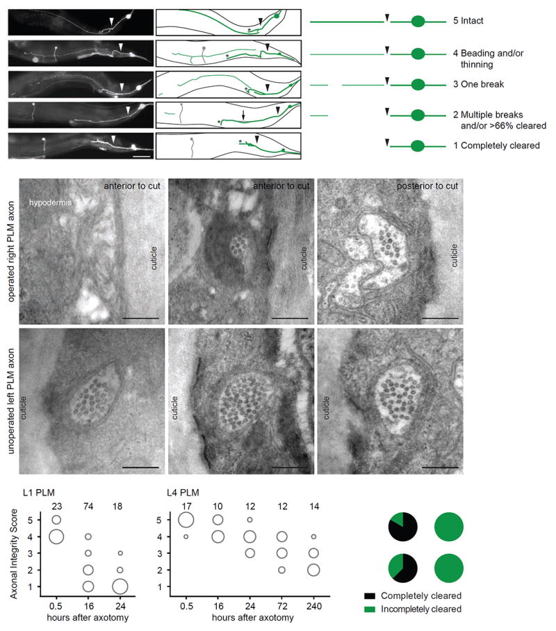
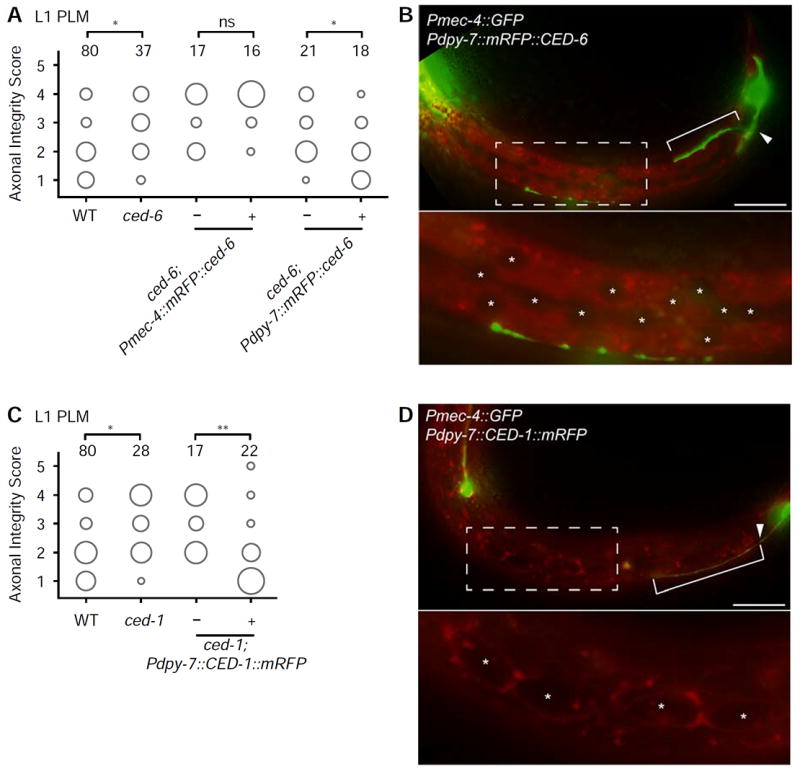
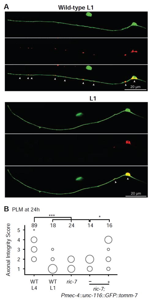
Axonal degeneration is a characteristic feature of neurodegenerative disease and nerve injury. Here, we characterize axonal degeneration in Caenorhabditis elegans neurons following laser-induced axotomy. We show that this process proceeds independently of the WLD(S) and Nmnat pathway and requires the axonal clearance machinery that includes the conserved transmembrane receptor CED-1/Draper, the adaptor protein CED-6, the guanine nucleotide exchange factor complex Crk/Mbc/dCed-12 (CED-2/CED-5/CED-12), and the small GTPase Rac1 (CED-10). We demonstrate that CED-1 and CED-6 function non-cell autonomously in the surrounding hypodermis, which we show acts as the engulfing tissue for the severed axon. Moreover, we establish a function in this process for CED-7, an ATP-binding cassette (ABC) transporter, and NRF-5, a lipid-binding protein, both associated with release of lipid-vesicles during apoptotic cell clearance. Thus, our results reveal the existence of a WLD(S)/Nmnat-independent axonal degeneration pathway, conservation of the axonal clearance machinery, and a function for CED-7 and NRF-5 in this process.


Figures

Figure 1
Figure 1. Characterization of axonal degeneration in C. elegans neurons following laser-induced axotomy
(A) Representative images and schematics of progressive stages of axonal degeneration after axotomy in PLM neurons in different L4 animals expressing the zdIs5(Pmec-4∷GFP) transgene. Arrowheads indicate location of axotomy, asterisks indicate the end of the regrowing axon, and arrow points to a distal fragment. The PVM neuron, which also expresses GFP, is represented in gray. Scale bar indicates 50 μm. Degeneration of the distal fragment classified as the Axonal Integrity Score with a value of 1 (completely cleared) to 5 (intact). The panel on the right is a schematic representation of these phenotypes. (B-G) Representative EM images of the degenerating PLM axon on the axotomized side (B-D) compared to the intact PLM axon on the non-operated side (E-G). The axon is intact upstream of the injury site (D), whereas in regions distal to the injury site it becomes severely reduced in diameter (C) or completely absent (B) (see also Figure S1E-S1F). On the contrary, on the unoperated contralateral side, PLM is intact along its entire length (E-G). (H,I) Axonal degeneration at multiple time points post-axotomy of the PLM neurons at L1 (H) or L4 (I) stages. The area of each circle represents the proportion of data points that fall into that category; n value indicated on top of each bubble graph. Each animal was imaged at only one time point. (J) Pie graph representing the complete clearance (score of 1) after 24 hours in PLM and DD neurons in L1 and L4 animals.
Figure 2
Figure 2. Overexpression of WldS or the endogenous Nmnat genes (nmat-1 and 2) has no protective effect against axonal degeneration after laser-induced axotomy in PLM neurons
(A) Axonal degeneration of representative transgenic strains expressing Pmec-4∷WldS at low (5 ng/μL), medium (10 ng/μL), or high (20 ng/μL) concentrations, 16 hours post-axotomy at the L1 stage. (B) Axonal degeneration of the PLM neuron in representative Pmec-4∷nmat-1 and Pmec-4∷nmat-2 lines at low (5 ng/μL) and high (20 ng/μL) concentrations 16 hours post-axotomy at the L1 stage. The high concentration of Pmec-4∷nmat-2 induced an increase in degeneration. (C) Quantification of PLM axonal degeneration at the L1 stage in rpm-1(ju41), dlk-1(ju476), tir-1(tm3036) or tir-1(ok1052) animals compared to wild-type (WT) animals 16 hours post-axotomy. The area of each circle represents the proportion of data points that fall into that category; n value indicated on top of each bubble graph. * p<0.05 as determined by either a Mann-Whitney test or a Kruskal-Wallis and Dunn’s test.
Figure 3
Figure 3. Conserved components of the apoptotic cell clearance machinery regulate the axonal degeneration phenotype of the PLM neurons
(A) Mutations in the apoptotic cell clearance genes ced-6(n1813), ced-1(e1735), ced-12(bz187), ced-10(n3246), ced-5(n1812), ced-2(e1752), ced-7(n2094), ced-7(n2690), and nrf-5(sa513) delay axonal degeneration in L1 animals. (B) Mutations in the other apoptotic-related genes ced-3(n2452), ced-4(n1162), ttr-52(sm211), psr-1(ok714), ina-1(gn144), tat-1(tm3117), and scrm-1(tm805) have no effect on axonal degeneration following axotomy in L1 animals. The area of each circle represents the proportion of data points that fall into that category; n value indicated on top of each bubble graph. * p<0.05, ** p<0.005, *** p<0.0005 as determined by a Kruskal-Wallis and Dunn’s or a Mann-Whitney test.
Figure 4
Figure 4. CED-1 and CED-6 function in the hypodermis
(A) Cell-specific expression of Pmec-4∷mRFP∷CED-6 indicates that the molecule does not act cell-autonomously in PLM neuron, whereas expression of Pdpy-7∷mRFP∷CED-6 in the hypodermis is sufficient to rescue the diminished degeneration and clearance in ced-6(n1813) mutants. This strain is representative for three independent rescue strains (data not shown). (B) Localization of Pdpy-7∷mRFP∷CED-6 in the hypodermis in an animal at the L1 stage, 16 hours after axotomy. (C) Cell-specific expression of Pdpy-7∷CED-1∷mRFP is sufficient to rescue the diminished degeneration and clearance in ced-1(e1735) mutants. This strain is representative of three independent rescue strains (data not shown). (D) Localization of Pdpy-7∷CED-1∷mRFP in the hypodermis and seam cells in an animal at the L1 stage, 16 hours after axotomy. The area of each circle in panels A and C represents the proportion of data points that fall into that category; n value indicated on top of each bubble graph. * p<0.05, ** p<0.005, as determined by a Kruskal-Wallis and Dunn’s or a Mann-Whitney test. Scale bars indicate 20 μm. Arrowheads indicate the location of axotomy, brackets indicate regrowth, and asterisks mark the hypodermal and seam nuclei.
Figure 5
Figure 5. Mitochondria are largely absent from the PLM axon in ric-7 mutant animals and are important for delaying axonal degeneration
(A) Mitochondria visualized through expression of TOMM-20∷mRFP at the L1-stage in wild-type (top panels) and ric-7(nu447) (bottom panels) animals, highlighting the lack of mitochondria within the axons of PLM in ric-7 mutants. (B) PLM axons of L4 ric-7(nu447) animals show a significant increase in degeneration and clearance compared to wild-type L4 animals after 24 hours, closely resembling the degree of degeneration and clearance in wild-type L1 animals. This defect can be rescued with cell-autonomous expression of UNC-116∷GFP∷TOMM-7 to restore mitochondrial localization in the axon. The area of each circle represents the proportion of data points that fall into that category; n value indicated on top of each bubble graph. * p<0.05, *** p<0.0005 as determined by a Kruskal-Wallis and Dunn’s or a Mann-Whitney test.

References

    1. Adalbert R, Gillingwater TH, Haley JE, Bridge K, Beirowski B, Berek L, Wagner D, Grumme D, Thomson D, Celik A, et al. A rat model of slow Wallerian degeneration (WldS) with improved preservation of neuromuscular synapses. Eur J Neurosci. 2005;21:271–277. - PubMed
    1. Avery MA, Rooney TM, Pandya JD, Wishart TM, Gillingwater TH, Geddes JW, Sullivan PG, Freeman MR. WldS prevents axon degeneration through increased mitochondrial flux and enhanced mitochondrial Ca2+ buffering. Curr Biol. 2012;22:596–600. - PMC - PubMed
    1. Avery MA, Sheehan AE, Kerr KS, Wang J, Freeman MR. WldS requires Nmnat1 enzymatic activity and N16-VCP interactions to suppress Wallerian degeneration. J Cell Biol. 2009;184:501–513. - PMC - PubMed
    1. Awasaki T, Tatsumi R, Takahashi K, Arai K, Nakanishi Y, Ueda R, Ito K. Essential role of the apoptotic cell engulfment genes draper and ced-6 in programmed axon pruning during Drosophila metamorphosis. Neuron. 2006;50:855–867. - PubMed
    1. Beirowski B, Babetto E, Coleman MP, Martin KR. The WldS gene delays axonal but not somatic degeneration in a rat glaucoma model. Eur J Neurosci. 2008;28:1166–1179. - PubMed

Publication types

MeSH terms